Best Ultrasound Scan in Hyderabad at Affordable Price
At PACE Hospitals, we provide advanced ultrasound scan in Hyderabad, Telangana using cutting-edge imaging technology operated by highly experienced radiologists. From abdominal ultrasound and pregnancy scans to pelvic, thyroid, breast, and Doppler ultrasound, we prioritize precision, patient safety, and diagnostic clarity. We specialize in 3D ultrasound scans in Hyderabad for detailed fetal imaging and 4D ultrasound scans in Hyderabad for real-time motion visuals, especially during pregnancy. Our services are designed to be affordable, with a transparent ultrasound scan price in Hyderabad, India for both scheduled and walk-in patients. For those seeking the best ultrasound in Hyderabad, PACE Hospitals ensures accurate results, fast reporting, and compassionate, patient-centered care — all in one place.
Book a USG Test - Ultrasonography
Ultrasound near me in Hyderabad Appointment
Ultrasonography / Ultrasound Scan / USG Test price in Hyderabad
Ultrasound / USG Imaging Test Preparation
Knowing how to prepare for your ultrasound scan in Hyderabad can help ensure accurate results and a smooth experience. At PACE Hospitals, our care team will guide you based on the type of ultrasound you’re scheduled for.
Ultrasound / USG Scan General Preparation Guidelines:
- Clothing: Wear loose, comfortable clothing. You might be asked to change into a hospital gown.
- Documents: Bring any previous scan reports, prescriptions, and your identification for verification.
- Arrival Time: Arrive 15–20 minutes before your scheduled appointment for registration and to receive pre-scan instructions.
Ultrasound / USG Test-Specific Preparations:
🟢 Abdominal Ultrasound:
- Fasting: Fast for 6–8 hours before the scan.
- Diet: Avoid carbonated drinks and fatty foods before the test.
- Water: Water may be allowed, but avoid eating anything.
🟢 Pelvic Ultrasound / Pregnancy Ultrasound:
- Hydration: Drink 3–4 glasses of water (approximately 1 liter) one hour before the scan.
- Urination: Do not urinate until after the scan is completed. A full bladder is necessary for a clearer view.
🟢 Thyroid, Breast, or Neck Ultrasound:
- Preparation: No special preparation is needed.
- Jewelry: Avoid wearing jewelry around the neck or chest area.
🟢 Doppler Ultrasound (Limbs, Abdomen, etc.):
- Fasting: May require fasting depending on the body part being examined. You will receive specific instructions.
- Clothing: Wear clothing that allows easy access to the area that will be scanned.
Important Note: Always follow the specific instructions given to you by your referring physician or the diagnostic team at PACE Hospitals. Incorrect preparation could lead to the need to reschedule your scan.
What is Ultrasound?
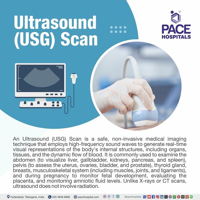
Ultrasound scan uses high-frequency sound waves to view inside the body. Because ultrasound pictures are collected in real time, they can reveal the movement of the body's internal organs as well as blood flow through the blood vessels. Unlike X-ray imaging, ultrasound imaging, does not expose the patient to ionising radiation.
In a USG test, an ultrasound probe (USG transducer) is positioned directly on the skin or within a bodily orifice (a body opening). A tiny layer of gel is applied to the skin to transmit USG waves from the transducer to the body.
The ultrasound scan images form from waves reflecting off internal structures. The sound signal’s intensity and the duration of its journey through the body provide the data needed to create the images.
Ultrasound is also called ultrasonography or sonography. Ultrasound images may be called sonograms. An ultrasound is usually done by a sonographer, a healthcare practitioner with special training to do ultrasound scans.
USG full form
The full form of USG in a medical context is Ultrasound Sonography. a powerful yet gentle imaging technique that’s been helping doctors "see" inside the body for decades.
USG meaning
The word "Ultrasound Sonography (USG)" is derived from the Latin words, "ultra" meaning beyond and sonus-meaning sound, and "graphy" meaning process of recording.
The technology advanced significantly over the twentieth century, becoming a cornerstone in medical diagnostics due to its capacity to generate real-time images without ionising radiation.
Historically, ultrasound technology began with static imaging modes such as A-mode and B-mode in the 1970s. It evolved to real-time scanning in 1979.
The development of Doppler ultrasound in the 1980s expanded its applications by allowing the visualisation of blood flow and movement. Ultrasound is now widely employed for a variety of applications, including foetal monitoring, identifying soft tissue diseases, and guiding medical therapies.
Types of Ultrasounds
Ultrasound technology is classified into several types, each designed for a distinct diagnostic purpose. Here are the major types of ultrasounds:
USG in pregnancy
An ultrasound pregnancy test, also known as a prenatal ultrasound, or obstetric ultrasound, or pregnancy ultrasound is an essential imaging procedure used to monitor the health and development of the foetus during pregnancy. The findings from these scans are documented in a USG report of pregnancy, which provides critical insights into foetal growth, maternal health, and overall pregnancy progression.
The 1st USG in pregnancy is usually performed during the first trimester, around 7 to 8 weeks after the last menstrual period. This initial ultrasound is crucial for confirming the pregnancy, determining the gestational age, detecting a fetal heartbeat, and identifying multiple pregnancies (e.g., twins or triplets). It also evaluates the uterus and ovaries to ensure there are no complications.
A pregnancy ultrasound schedule typically includes several key scans throughout gestation to monitor the health and development of both the mother and the fetus, assess placental position, check amniotic fluid levels, and screen for abnormalities.
Each scan plays an important role in ensuring a healthy pregnancy and is reflected in the USG report of pregnancy, which serves as a comprehensive record for healthcare professionals to guide prenatal care effectively.
USG abdomen
A whole abdomen ultrasound, also called as an ultrasound scan abdomen or USG whole abdomen, is a non-invasive imaging test widely used to evaluate the organs and structures within the abdominal cavity. This includes the liver, kidneys, pancreas, spleen, gallbladder, and major blood vessels.
The USG abdomen test is particularly effective in diagnosing conditions such as abdominal pain, fluid accumulation (ascites),
kidney stones,
gallstones, and tumors. Additionally, a specialized USG abdomen and pelvis scan can provide detailed insights into the pelvic organs, including the bladder and reproductive organs. By offering real-time imaging, this procedure helps healthcare experts identify abnormalities and plan appropriate treatments efficiently.
Pelvic ultrasound
A pelvic ultrasound is a non-invasive imaging test that uses sound waves to create pictures of the organs in the pelvis. It's also known as pelvic sonography or pelvic scan.
The results of a pelvic ultrasound are documented in a pelvic ultrasound report, which outlines the findings regarding organ size, shape, and any detected abnormalities. This report is essential for guiding further medical evaluation or treatment.
While pelvic ultrasounds are generally safe and non-invasive, patients may wonder about potential pelvic ultrasound side effects. The procedure does not involve radiation and is considered very safe; however, mild discomfort may occur during a pelvic transvaginal ultrasound due to probe insertion. Overall, pelvic ultrasounds are invaluable tools for diagnosing and managing gynaecological and urological conditions effectively.
Transvaginal ultrasound
A transvaginal ultrasound scan is a specialized imaging procedure that uses high-frequency sound waves to create detailed images of pelvic organs, including the uterus, ovaries, fallopian tubes, and cervix. It provides clearer images than abdominal ultrasounds due to the proximity of the transducer to the pelvic structures.
The transvaginal ultrasound protocol involves inserting a lubricated transducer, covered with a protective sheath, into the vagina after the patient empties the bladder. This allows for optimal visualization of the pelvic organs. The procedure, which typically takes 15 to 30 minutes, is minimally invasive and well-tolerated.
Transvaginal ultrasounds are commonly used to evaluate pelvic pain, abnormal bleeding, infertility issues, and to monitor early pregnancy. The results are compiled into a detailed report to assist healthcare experts in diagnosing conditions and planning treatments effectively.
Breast ultrasound
A breast ultrasound is a non-invasive imaging test that uses high-frequency sound waves to create detailed images of breast tissue. It is often used to evaluate abnormalities such as lumps or changes detected during a physical exam or mammogram. This test is particularly helpful in distinguishing between fluid-filled cysts and solid masses, which may require further investigation for. A breast cancer ultrasound can provide valuable insights into suspicious areas and assist in guiding biopsies for accurate diagnosis.
During the breast ultrasound procedure, the patient lies on an examination table after undressing from the waist up. A clear, water-based gel is applied to the skin, allowing sound waves to transmit effectively. A handheld transducer is moved over the breast to capture images, which are displayed on a monitor. The procedure typically lasts 15–30 minutes, is painless, and does not involve radiation, making it safe for all patients.
Breast ultrasounds are often performed as a supplemental screening tool alongside mammograms, especially for individuals with dense breast tissue or those at high risk for breast cancer. The results are interpreted by a radiologist and compiled into a report, guiding further evaluation or treatment if necessary. This imaging test plays a crucial role in breast cancer detection and management, offering detailed insights for healthcare professionals.
Doppler ultrasound
Doppler ultrasound is a non-invasive imaging technique that uses sound waves to assess blood flow in vessels and organs. Among its specialized forms, color doppler ultrasound provides real-time, color-coded visuals of blood flow direction and velocity, making it essential for diagnosing vascular conditions.
In pregnancy, colour doppler ultrasound is widely used to monitor fetal health by evaluating blood flow in the placenta and umbilical cord, ensuring proper oxygen and nutrient delivery to the fetus. Additionally, renal doppler ultrasound focuses on the kidneys, helping to detect issues like renal artery stenosis or impaired blood flow, which are important for diagnosing and managing kidney-related diseases. This versatile tool is integral in both prenatal care and vascular diagnostics.
Renal ultrasound
Renal ultrasound is a non-invasive imaging technique widely used to assess the kidneys, ureters, and bladder for various conditions. The renal ultrasound protocol involves scanning the patient in supine or lateral positions using a curvilinear transducer to capture detailed images of renal anatomy.
Common renal ultrasound indications include evaluating flank pain, haematuria, urinary tract obstruction, kidney stones, cysts, tumors, and infections. It is also used to monitor chronic kidney disease (CKD) progression or assist in procedures like renal biopsies. A normal renal ultrasound generally shows kidneys with normal size and shape, homogeneous echotexture, distinct corticomedullary differentiation, and a central hypoechoic renal pelvis. This safe and effective modality is crucial for diagnosing and managing renal disorders.
Thyroid ultrasound
Thyroid ultrasound is a vital diagnostic tool for evaluating thyroid disorders, including thyroiditis and thyroid nodules. In cases of thyroiditis, particularly Hashimoto's thyroiditis, ultrasound typically reveals an enlarged, hypoechoic gland with a heterogeneous echotexture and micronodules surrounded by echogenic septations, indicative of lymphocytic infiltration. On color Doppler imaging, vascularity can vary from slight to marked hypervascularity, often associated with hypothyroidism.
For thyroid nodules, ultrasound typically shows them as hypoechoic with thin hypoechoic halos, smooth or irregular margins, and variable vascularity. These characteristics aid in differentiating between benign and malignant lesions, making this imaging modality crucial for diagnosing thyroid conditions and guiding further management, such as biopsies or treatment planning.
Scrotal ultrasound
Scrotal ultrasound is an important imaging technique used to evaluate various scrotal conditions, including the assessment of testicular abnormalities. A scrotal doppler ultrasound is often employed to assess blood flow within the testicles and surrounding structures, which is particularly important in cases of acute scrotal pain or suspected torsion. The findings from this examination are documented in a scrotal ultrasound report, which includes detailed descriptions of the testicular morphology, size, echogenicity, and any pathological lesions identified.
This comprehensive report aids healthcare professionals in diagnosing conditions such as epididymitis, varicoceles, and tumors, ensuring appropriate management and follow-up for patients.
Musculoskeletal ultrasound
Musculoskeletal ultrasound is a rapidly evolving imaging technique that provides valuable insights into various musculoskeletal disorders. Understanding the basics of musculoskeletal ultrasound involves recognizing its ability to visualize muscles, tendons, ligaments, and joints in real-time using high-frequency sound waves.
This non-invasive method is particularly advantageous due to its accessibility, quick scan times, and lack of ionizing radiation. It allows for dynamic examinations, enabling clinicians to assess conditions such as sprains, tears, and entrapments effectively. By mastering the fundamentals of this imaging modality, healthcare providers can enhance their diagnostic capabilities and improve patient outcomes in musculoskeletal care.
3D ultrasound
A 3D ultrasound scan is an advanced imaging technique that provides three-dimensional images of internal structures, allowing for a more comprehensive view compared to traditional 2D ultrasound. This method is particularly beneficial in obstetrics, as it produces 3D ultrasound pictures that can help visualize fetal anatomy and detect congenital anomalies more effectively. However, like any medical procedure, there are 3D ultrasound pros and cons to consider.
The advantages include improved diagnostic accuracy and enhanced visualization of complex structures, while the drawbacks may involve higher costs and the potential for overdiagnosis due to the detailed images produced. Overall, 3D ultrasound represents a significant advancement in imaging technology, offering valuable insights for both clinicians and patients.
4D ultrasound
4D ultrasound is an advanced imaging technique that enhances traditional ultrasound by adding the dimension of time, allowing for real-time visualization of moving structures within the body. This technology utilizes high-frequency sound waves to create three-dimensional images, which are then compiled into a live video format.
It is particularly popular in obstetrics, as it enables expectant parents to observe their unborn child's movements, such as yawning or stretching, providing a unique and emotional connection before birth. Additionally, 4D ultrasound can assist healthcare professionals in monitoring fetal development and identifying potential abnormalities, making it a valuable tool in prenatal care.
Transesophageal echocardiogram
A transesophageal echocardiogram is an advanced imaging procedure that provides detailed views of the heart by utilizing a specialized probe inserted into the oesophagus. This approach allows for clearer images than traditional methods, as it positions the transducer closer to the heart, minimizing interference from surrounding tissues.
The procedure typically involves mild sedation to ensure patient comfort, along with local anaesthetic to numb the throat. During the examination, high-frequency sound waves are emitted, creating real-time images that help healthcare professionals assess heart structures, detect abnormalities, and evaluate conditions such as valve disease or blood clots. The insights gained from this imaging technique are invaluable for diagnosing and managing various cardiac issues effectively.
Types of Ultrasounds in Pregnancy
Types of ultrasound scans in pregnancy include 2D, 3D, and 4D scans, as well as transvaginal and transabdominal scans, each serving different purposes, from confirming pregnancy and fetal development to assessing fetal health and heart function.

Ultrasound Indications
Ultrasound is a widely utilized imaging modality with a broad range of indications across medical specialties. Its ability to provide real-time, non-invasive visualization of internal structures makes it an essential diagnostic and therapeutic tool. The indications for ultrasound extend from routine evaluations to critical assessments in emergency and specialized care settings.
Ultrasound, as a diagnostic modality, is predominantly used in:
- Obstetric and gynecologic applications
- Abdominal imaging uses
- Cardiac assessment techniques
- Vascular health evaluation
- Musculoskeletal imaging
- Pediatric diagnostic applications
- Thyroid and breast imaging techniques
- Emergency medicine utilization
- Interventional procedures guidance
Obstetric and gynecologic applications
In obstetrics, ultrasound is indicated for monitoring fetal development, determining gestational age, assessing placental health, and detecting congenital anomalies (abnormalities present at birth). It is also used to evaluate multiple pregnancies and fetal positioning before delivery. In gynecology, ultrasound is indicated for diagnosing ovarian cysts, uterine fibroids, endometrial abnormalities, and ectopic pregnancies.
Abdominal imaging uses
Ultrasound is commonly indicated for evaluating abdominal organs such as the liver, gallbladder, pancreas, kidneys, and spleen. It is used to diagnose conditions like gallstones, kidney stones, liver cirrhosis, and abdominal masses. Additionally, ultrasound guides interventional procedures like biopsies or the aspiration of fluid collections.
Cardiac assessment techniques
In cardiology, echocardiography is indicated for assessing heart structure and function. It is used to diagnose valve disorders, cardiomyopathies, congenital heart defects, and pericardial effusion. Doppler ultrasound is specifically indicated for evaluating blood flow within the heart and major vessels.
Vascular health evaluation
Vascular ultrasound is indicated for assessing blood flow in arteries and veins. It helps diagnose conditions such as deep vein thrombosis (DVT), arterial stenosis, aneurysms, and varicose veins. These indications are crucial for planning interventions like vascular or angioplasty surgery.
Musculoskeletal imaging
Ultrasound is indicated for evaluating musculoskeletal conditions such as tendon injuries, ligament tears, muscle strains, joint effusions, and soft tissue masses. It is also used to guide therapeutic procedures like corticosteroid injections or the aspiration of fluid from joints.
Pediatric diagnostic applications
In pediatrics, ultrasound is indicated for assessing neonatal brain abnormalities such as hydrocephalus or intracranial hemorrhage. It is also used to evaluate hip dysplasia in infants and monitor kidney function in pediatric patients.
Thyroid and breast imaging techniques
Ultrasound is indicated for assessing thyroid nodules, goiters, or other thyroid abnormalities. In breast imaging, it complements mammography by evaluating masses or guiding biopsies to rule out malignancy.
Emergency medicine utilization
In emergency medicine, ultrasound is indicated for rapid assessment of trauma patients, using techniques like Focused Assessment with Sonography for Trauma (FAST) to identify internal bleeding or organ injuries. It is also used to evaluate acute conditions such as ectopic pregnancy or vascular emergencies.
Interventional procedures guidance
Ultrasound plays a significant role in guiding various interventional procedures due to its real-time imaging capabilities. It is indicated for performing fine-needle aspirations and biopsies of soft tissue masses or fluid collections with high accuracy. Common procedures that utilize ultrasound guidance include:
- Thoracentesis: Draining fluid from the pleural space.
- Paracentesis: Removing fluid from the abdominal cavity.
- Cyst aspiration: Draining cysts or abscesses.
- Joint injections: Administering corticosteroids or anesthetics into joints.
- Biopsies: Guiding needle placement for tissue sampling.
- Foreign body retrieval: Locating and removing foreign objects embedded in soft tissues.
- Peripheral nerve hydrodissection: Relieving nerve entrapment by injecting fluid around the nerve.
Emergency Ultrasound Indications
Emergency ultrasound is an indispensable diagnostic tool in acute care settings, offering rapid, real-time imaging that aids in the management of life-threatening conditions. It is widely used for Focused Assessment with Sonography in Trauma (FAST) to detect internal bleeding in trauma patients, enabling timely surgical intervention. In obstetrics, emergency ultrasound plays a critical role in diagnosing ectopic pregnancies, preventing severe complications.
It is also valuable for evaluating cardiac conditions such as pericardial effusion and thoracic issues like pneumothorax or pleural effusions.
Additionally, ultrasound is utilized to assess abdominal emergencies such as gallbladder disease, appendicitis, or obstructive uropathy, as well as vascular conditions like deep vein thrombosis (DVT). Beyond diagnostics, emergency ultrasound is integral for guiding procedures such as central line placement, thoracentesis, and paracentesis with precision and safety. Its versatility and efficiency make it a cornerstone of modern emergency medicine, improving patient outcomes in critical situations.

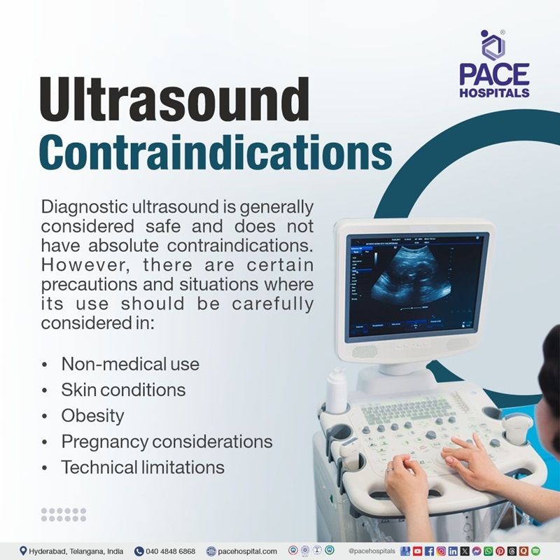
Ultrasound Contraindications
Diagnostic ultrasound is generally considered safe and does not have absolute contraindications. However, there are certain precautions and situations where its use should be carefully considered in:
- Non-medical use: Ultrasound should not be used for non-medical purposes, such as obtaining pictures of the fetus solely for entertainment or determining fetal sex without a medical reason.
- Skin conditions: Ultrasound may be difficult or inappropriate if there is skin sepsis or lack of access preventing skin/transducer contact.
- Obesity: In obese patients, the thick subcutaneous fat layer can limit sound wave penetration, making alternative imaging methods like
CT scans more suitable for deep structures.
- Pregnancy considerations: While ultrasound is safe during pregnancy, it should only be performed when medically indicated to minimize unnecessary exposure.
- Technical limitations: Ultrasound waves are also disrupted by air or gas, limiting the ability to image organs obscured by air-filled structures like the bowel (intestines).
Overall, diagnostic ultrasound is a valuable tool with no absolute contraindications, but it should be used judiciously and only when there is a medical benefit.
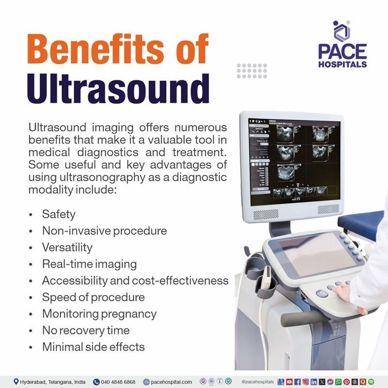
Benefits of Ultrasound
Ultrasound imaging offers numerous benefits that make it a valuable tool in medical diagnostics and treatment. Some useful and key advantages of using ultrasonography as a diagnostic modality include:
- Safety
- Non-invasive procedure
- Versatility
- Real-time imaging
- Accessibility and cost-effectiveness
- Speed of procedure
- Monitoring pregnancy
- No recovery time
- Minimal side effects
Safety
Unlike X-rays and CT scans, ultrasound uses sound waves instead of ionizing radiation, significantly reducing the risk of harmful effects on patients.
Non-invasive procedure
Ultrasound is a non-invasive technique that does not require needles or incisions, making it painless and suitable for a wide range of patients, including pregnant women.
Versatility
Ultrasound excels at visualizing soft tissues, which are not clearly seen on X-rays. This capability is crucial for diagnosing conditions in organs such as the liver, kidneys, and reproductive organs.
Real-time imaging
Ultrasound provides real-time imaging, allowing for immediate assessment of moving structures, which is particularly useful during procedures like biopsies or fluid aspirations.
Accessibility and cost-effectiveness
Ultrasound machines are more portable and less expensive than other imaging modalities, making them accessible in various healthcare settings.
Speed of procedure
Ultrasound examinations can often be completed within minutes to an hour, facilitating faster diagnosis and treatment decisions.
Monitoring pregnancy
Ultrasound is extensively used in obstetrics to monitor fetal development and detect potential abnormalities during pregnancy.
No recovery time
Patients generally experience no downtime after an ultrasound, allowing them to resume normal activities immediately.
Minimal side effects
There are virtually no known harmful side effects when ultrasound is performed correctly, making it a safe option for diagnostic imaging.
These benefits underscore the importance of ultrasound as a diagnostic tool across various medical fields, enhancing patient care while minimizing risks.

Ultrasonography Procedure
Ultrasound imaging serves as a fundamental component of modern diagnostics, offering real-time visualization of soft tissues, organs, and blood flow without the use of ionizing radiation. A standardized procedure for conducting an ultrasound involves patient preparation, selecting the appropriate transducer, and capturing images.
Ultrasound procedure steps:
Preparation
Patients may be asked to remove clothing or jewellery from the area being examined.
Positioning
The patient will lie down on an examination table, usually on their back, but may be asked to turn to either side depending on the area being examined.
Application of gel
A water-based gel is applied to the skin over the area of interest. This gel helps eliminate air pockets that can block sound waves and ensures better contact between the transducer and the skin.
Transducer placement
The sonographer (ultrasound technician) will use a handheld device called a transducer. The transducer is pressed against the gel-covered skin and moved around to capture images of the internal structures.
Image acquisition
The transducer emits high-frequency sound waves into the body. These sound waves bounce off organs and tissues, returning echoes that are captured by the transducer. The echoes are processed by a computer to create real-time images displayed on a monitor. The sonographer may capture still images or video loops as needed.
Adjustments
The sonographer may ask the patient to change positions or hold their breath briefly to improve image quality or to access different areas.
Completion of the exam
Once sufficient images have been obtained, the procedure is concluded. The gel is wiped off the patient's skin, and they can usually resume normal activities immediately.
Post-procedure follow-up
The images will be reviewed by a radiologist or physician who will interpret the results and discuss any findings with the patient, potentially scheduling follow-up appointments if needed.
Ultrasonography is generally considered a safe and effective imaging technique, but it can have complications, particularly when used for interventional procedures. Here are some potential complications associated with ultrasonography.
Principle of Ultrasound
Ultrasound imaging relies on the "pulse-echo" principle, where a transducer emits sound waves that bounce off internal structures, and the returning echoes are processed to create an image.
The mechanism of the ultrasound procedure is based on:
- Sound wave generation: An ultrasound machine generates high-frequency sound waves using a transducer, which is a device that converts electrical energy into mechanical energy (sound waves).
- Transmission: These sound waves are transmitted into the body through a coupling gel, which helps to ensure good contact between the transducer and the skin.
- Reflection and scattering: As the sound waves travel through the body, they encounter different tissues and organs, which have varying densities and acoustic properties. When the sound waves hit these interfaces, they are reflected towards the transducer.
- Echo reception: The transducer also acts as a receiver, capturing the returning echoes.
- Image formation: The ultrasound machine processes the received echoes to create an image, where different tissues and structures are displayed based on their ability to reflect sound waves.
- Doppler effect: In some ultrasound applications, the Doppler effect is used to measure the flow of blood or other fluids.
- Attenuation: As sound waves travel through the body, they lose energy due to absorption and scattering, a phenomenon known as attenuation.
- Time-gain compensation:
This is used to amplify signals which have taken longer to return, which amplifies signals returned from deep tissues.
Ultrasound Complications
Diagnostic ultrasonography is generally considered safe, but it can have potential complications and risks, particularly related to thermal and mechanical effects on tissues. The diagnostic ultrasonography complications may include:
- Localized pain or discomfort
- Cavitation and heating effects
- Pregnancy imaging risks
- Thermal effects
- Radiation pressure
- Misdiagnosis
Localized pain or discomfort
While the ultrasound procedure itself is generally painless, patients may experience discomfort if pressure is applied to sensitive areas or if internal probes are used.
Cavitation and heating effects
Ultrasound waves can cause slight heating of tissues and, in rare cases, produce small pockets of gas (cavitation) in body fluids or tissues. The long-term consequences of these effects are not fully understood.
Pregnancy imaging risks
Some studies have reported potential effects of ultrasound exposure during pregnancy, such as low birth weight or delayed speech, but there is no conclusive evidence of a causal relationship.
Thermal effects
Ultrasound can cause a slight increase in tissue temperature, but this is typically not harmful in diagnostic settings. However, prolonged exposure should be avoided, especially in sensitive areas like the fetus during pregnancy.
Radiation pressure
Although the severity of radiation pressure as a hazard is low, it is always present. Little is known about any associated cell responses, making it difficult to evaluate the risk.
Misdiagnosis
Operator error or equipment limitations can lead to misdiagnosis, which is a significant complication of ultrasound use. Proper training and adherence to safety guidelines are crucial to minimize this risk.
While these complications are relatively uncommon, they underscore the importance of conducting ultrasound procedures with care and by trained professionals to minimize risks and ensure patient safety.
Questions that the patients can ask the healthcare team about ultrasonography?
Patients can ask the healthcare team the following questions about ultrasonography to better understand the procedure and its implications:
- What is the purpose of this ultrasound?
- What conditions or abnormalities can this ultrasound detect?
- Are there any risks or complications associated with this procedure?
- How should I prepare for the ultrasound?
- Should I wear specific clothing or bring anything to the appointment?
- What happens during the ultrasound procedure?
- Will it be external or involve inserting a transducer into a body cavity?
- How long will the procedure take?
- Will I experience any discomfort during the procedure?
- When and how will I receive my results?
- If abnormalities are found, what additional tests or treatments might be needed?
- How often should I undergo this type of ultrasound based on my condition?
These questions can help patients feel informed and prepared for their ultrasound exam.
Difference between Sonogram and Ultrasound
sonogram vs ultrasound
The terms sonogram and ultrasound are closely related but refer to distinct aspects of the imaging process, often leading to confusion. The table below contains the key differences between sonogram and the ultrasound:
| Aspect | Sonogram | Ultrasound |
|---|---|---|
| Definition | The visual image or record produced by the ultrasound process. | A diagnostic technique using high-frequency sound waves to create images. |
| Function | Displays the actual image produced by the ultrasound. | Captures sound waves that bounce off the body's tissues to create an image. |
| Role | The result or outcome (image) generated from the ultrasound. | A medical procedure or tool used to generate images. |
| Physical form | A visual or graphical representation of the data from the ultrasound. | Refers to the procedure or process, not the image itself. |
| Usage | Used for review and interpretation of the images captured. | Used for real-time imaging during a procedure or examination. |
| Example | The image of the fetus seen on a monitor is a sonogram. | A doctor performs an ultrasound to monitor a fetus during pregnancy. |
Difference between Ultrasound and CT scan
Ultrasound vs CT scan
Ultrasound and CT scans are both diagnostic imaging modalities used in medicine, although they work on different principles and serve different objectives. Here are the main distinctions between them:
| Aspect | Ultrasound | CT scan |
|---|---|---|
| Technology | Uses sound waves to create images. | Uses X-rays and computer processing for images. |
| Procedure | Non-invasive, no radiation, with a probe. | Involves X-ray exposure and a rotating machine. |
| Image quality | 2D, real-time images, good for soft tissues. | High-resolution, 3D images, detailed for bones and organs. |
| Radiation | No radiation | Involves X-ray radiation. |
| Uses | Pregnancy, organ exams, guiding procedures | Detects tumours, fractures, internal bleeding. |
Difference between Infrasound and Ultrasound
Infrasound and ultrasound represent the extremes of the acoustic spectrum, differing primarily in frequency and applications. Infrasound consists of sound waves below 20 Hz, often used for monitoring natural phenomena like earthquakes and animal communication. Ultrasound, with frequencies above 20 kHz, is widely used in medical imaging, therapy, and industrial applications such as cleaning and non-destructive testing
Frequently Asked Questions (FAQs) on Ultrasonography / Ultrasound Scan / USG Test
Is USG safe?
Ultrasound is considered safe as it uses non-ionizing sound waves rather than radiation. No significant adverse effects have been reported with diagnostic use, making it suitable even for sensitive populations like pregnant individuals.
Will ultrasound show inflammation?
Yes, inflammation can be detected on an ultrasound by observing thickened organ walls (e.g., bowel wall), fluid accumulation, or increased blood flow using Doppler settings. These findings help diagnose inflammatory conditions accurately.
Does ultrasound detect ectopic pregnancy?
Yes, transvaginal ultrasounds are highly effective in diagnosing ectopic pregnancies by identifying gestational sacs outside the uterus or associated signs like free fluid in the pelvis. Early detection prevents complications like rupture.
Does ultrasound require fasting?
Fasting is required for certain ultrasounds (e.g., abdominal) to reduce gas interference and ensure better visualization of organs like the gallbladder and pancreas. However, fasting is not needed for all types of ultrasounds.
What diseases can be detected by ultrasound?
Ultrasound can detect a wide range of diseases and conditions, including those affecting the reproductive system, abdominal organs, blood vessels, and musculoskeletal system, as well as being used during pregnancy to monitor fetal development.
What is TIFFA ultrasound?
TIFFA ultrasound (Targeted Imaging for Fetal Anomalies) is a detailed scan performed between 18 and 22 weeks of pregnancy to assess fetal anatomy and detect congenital anomalies. It evaluates key structures like the heart, brain, spine, and organs, ensuring early identification of abnormalities for timely intervention.
How many ultrasounds are done during pregnancy?
During a normal pregnancy, most women have two to three ultrasounds. However, the amount and timing may differ depending on factors such as the pregnant woman's health, pregnancy risks, and the gynecologist recommendations.
Can ultrasound detect intestinal problems?
Yes, ultrasound can detect intestinal problems such as bowel obstruction, Crohn's disease, and diverticulitis. It identifies abnormalities like bowel wall thickening, strictures, and abscesses. Doppler techniques enhance its accuracy by assessing vascular changes associated with inflammation, making it a valuable tool for non-invasive diagnostics in gastrointestinal disorders.
Can ultrasound detect intestinal cancer?
Ultrasound can detect intestinal cancer by identifying masses or abnormal bowel wall thickening. However, its sensitivity varies based on tumor location and operator expertise. Advanced imaging methods like CT or MRI are preferred for definitive diagnosis due to their ability to evaluate deeper structures and provide detailed visualization.
Can ultrasound detect liver problems?
Ultrasound is highly effective for detecting liver problems such as
fatty liver disease, cirrhosis, tumors, and bile duct obstructions. It evaluates liver size, structure, echogenicity, and blood flow patterns. Doppler imaging enhances the detection of vascular abnormalities associated with liver diseases.
What is TVS ultrasound?
Transvaginal Sonography (TVS) involves inserting a probe into the vagina to obtain detailed images of pelvic organs such as the uterus and ovaries. It is commonly used for gynecological assessments and early pregnancy evaluations due to its high-resolution imaging capabilities and ability to detect small abnormalities.
Why is gender determination via ultrasound illegal in India?
In India, revealing the gender of a baby via ultrasound (USG) is illegal under the Pre-Conception and Pre-Natal Diagnostic Techniques (PCPNDT) Act, aimed at preventing sex-selective abortions.
Can ultrasound detect cancer?
Ultrasound can identify suspicious masses in organs like the liver, thyroid, or breast that may indicate cancer. Doppler imaging assesses abnormal blood flow patterns in tumors. However, biopsy or advanced imaging techniques like MRI are required for confirming malignancy.
Can ultrasound detect pregnancy at 1 week?
No, ultrasound cannot detect pregnancy at 1 week since the gestational sac is not yet visible. Detection typically occurs around 4-5 weeks when hCG levels are elevated enough to visualize early pregnancy structures like the gestational sac.
Can ultrasound detect stomach cancer?
Ultrasound is not ideal for detecting stomach cancer due to limited visualization of deeper gastric structures. Endoscopic ultrasound (EUS) is preferred for assessing stomach wall layers and nearby lymph nodes, with higher accuracy in diagnosing malignancies.
Is sonography and ultrasound the same?
Yes, sonography and ultrasound refer to the same imaging technique that uses high-frequency sound waves to create images of internal organs. The terms are interchangeable in medical practice and are widely used for diagnostic purposes.
Would drinking alcohol before an ultrasound affect results?
Drinking alcohol before an abdominal ultrasound may affect results by altering liver function or causing gas accumulation in the intestines. These factors can interfere with image clarity and reduce diagnostic accuracy.
Would ovarian cancer show on ultrasound?
While ultrasound can detect masses in the ovaries, it cannot definitively diagnose ovarian cancer and further tests, like a biopsy, are needed to confirm if a mass is cancerous.
Why do we need to drink more water for a pelvic ultrasound?
A full bladder helps lift pelvic organs into view and acts as an acoustic window for better visualization during a pelvic ultrasound. This improves image clarity and diagnostic accuracy significantly.
Why is water needed for an ultrasound?
Water reduces air interference between the transducer and internal organs while distending certain structures like the bladder for better visualization. It enhances image quality significantly during abdominal or pelvic ultrasounds.
Why should we limit the use of ultrasound in pregnancy?
Although considered safe, limiting unnecessary ultrasounds during pregnancy minimizes potential risks like tissue heating or cavitation due to prolonged exposure to sound waves. Ethical guidelines recommend using ultrasounds only when medically necessary.
What is the difference between ultrasound and ultrasound therapy?
Diagnostic ultrasounds create images of internal structures using sound waves. Ultrasound therapy uses high-frequency sound waves therapeutically to treat conditions like muscle pain or inflammation by promoting tissue healing through mechanical vibrations.
Can ultrasound detect appendicitis?
Yes, appendicitis can be diagnosed via ultrasound by identifying an enlarged appendix (>6 mm), free fluid, or increased blood flow around it using Doppler imaging techniques. It is highly effective when performed by experienced operators.
Can ultrasound detect hernia?
Yes, hernias can be detected via ultrasound by visualizing abnormal protrusions of tissues through weak muscle walls. Doppler imaging may also assess blood flow in cases of strangulation or complications such as ischemia.
Is TVS ultrasound painful?
A transvaginal ultrasound (TVS) typically causes mild discomfort rather than pain. The procedure involves inserting a lubricated probe into the vagina, which may result in some pressure. However, this discomfort is usually minimal and temporary, resolving once the procedure is completed.
The probe is designed to fit comfortably, and techniques like deep breathing can help reduce any discomfort experienced during the scan. Overall, while some women may feel slightly uncomfortable, the procedure is generally well-tolerated and not painful.
What is level 2 ultrasound?
A Level 2 ultrasound, also known as a fetal anatomical survey or anomaly scan, is a detailed ultrasound performed during the second trimester (typically between 18 and 22 weeks) to assess fetal development and identify potential abnormalities.
What is USG KUB?
USG KUB, or ultrasound of the Kidneys, Ureters, and Bladder, is a non-invasive imaging technique that uses sound waves to create images of the urinary tract, helping diagnose conditions like kidney stones, infections, or structural abnormalities.
What is USG guided biopsy?
An ultrasound-guided biopsy is a procedure that uses ultrasound to guide a needle to remove a tissue sample for examination. It's a non-invasive diagnostic test that can help diagnose abnormalities like infection, inflammation, or malignancy.
What is banana sign on USG?
The banana sign on a fetal ultrasound refers to an abnormal appearance of the cerebellum, appearing curved or banana-shaped due to caudal displacement, and is often associated with open neural tube defects like spina bifida (a birth defect where the spine doesn’t fully close, potentially exposing the spinal cord and causing nerve issues).
What is lamda sign in USG?
The "lambda sign" or "twin peak sign" in ultrasound (USG) refers to a triangular projection of placental tissue extending between the layers of the intertwin membrane, strongly suggesting a dichorionic diamniotic (DCDA) twin pregnancy.
What are the different types of ultrasound probes?
Ultrasound probes vary significantly in design and application to suit diverse clinical needs. Common types include linear probes, ideal for imaging superficial structures like vessels and tendons, and convex probes, which provide a wider field of view for deeper organs such as the abdomen.
Phased array probes are compact and used for cardiac imaging, while endocavitary probes are designed for internal examinations like gynecological scans. Specialized probes like transesophageal echocardiography (TEE) probes offer detailed imaging of the heart via the esophagus, and pencil Doppler probes focus on vascular flow assessments. Advanced models, such as matrix probes, enable real-time volumetric imaging for complex diagnoses.
What is POCUS ultrasound?
Point-of-care ultrasound (POCUS) is the use of portable ultrasound machines by trained medical professionals to perform rapid, focused ultrasound scans at the patient's bedside or point of care, enhancing clinical decision-making and potentially expediting diagnosis and treatment.


