
Best Cancer & Oncology Hospital in Hyderabad | Expert Cancer Care
PACE Hospitals is one of the Best Cancer Hospitals in Hyderabad, Telangana, India, offering comprehensive cancer treatment tailored to patient needs.
The experienced and skilled multidisciplinary team of medical oncologists, surgical oncologists, and radiologists uses multimodal cancer treatment therapy like chemotherapy, radiation therapy, hormonal therapy, palliative care, targeted therapy and localized therapy to manage and treat all kinds of cancer, from solid tumours to blood cancer. The expert team of oncology doctor have vast expertise in the treatment of cancer, including:
- Stomach Cancer, Esophageal cancer, Colorectal cancer
- Head and Neck cancer, Oral cancer, Thyroid cancer
- Cervical cancer, Uterine Cancer, Ovarian Cancer, Breast Cancer
- Lung cancer, Leukaemia, Myeloma, Lymphoma
- Liver Cancer, Pancreatic cancer, Gallbladder Cancer
- Bladder cancer, Prostate cancer, Kidney cancer
Book an Appointment for Cancer Treatment
Cancer Treatment Appointment Enquiry
Why Choose PACE Hospitals for Cancer Care?
Comprehensive Cancer Care
Providing multimodal cancer treatment like chemotherapy, radiation therapy, targeted therapy, and localized therapy (surgeries) to a wide range of cancers from solid benign & malignant tumours to blood and lymphatic system cancer based on patient medical condition.
Advanced State-of-the-art Facility
Equipped with advanced and latest diagnostic equipments for precise screening of cancer staging & grading, robotic and minimally invasive surgical facilities for advanced cancer treatment with optimal outcome and high success rate.
Skilled & Experienced Oncology Doctors
A team of experienced cancer specialists, medical oncologists and surgical oncologists with vast experience in managing all kinds of cancer through medical or surgical approaches to eliminate cancer, relieving symptoms and reducing the risk of cancer, based on diagnosis and evaluation.
Leading Oncology Centre for Cancer Treatment in Hyderabad, Telangana
PACE Hospitals is one of the Best Oncology Hospitals in Hyderabad, India, committed to catering to holistic, precise, advanced treatments and unwavering patient support to fight against cancer. The team of medical oncology doctors, surgical oncology doctors, and highly skilled radiologists specializes in the treatment of all kinds of cancers, including head & neck cancers, GI cancers, gynaecological cancers, haematological malignancies with advanced cancer treatment modalities like minimally invasive surgical oncology, chemotherapy, targeted therapies, immunotherapy, and radiation oncology based on the nature and stages of the cancer.
The Department of Oncology at PACE Hospitals is equipped with state-of-the-art & advanced diagnostic facilities to ensure early diagnosis, customized treatment plans, and seamless patient care, leading the Fight Against Cancer with
Advanced Cancer Treatment in Hyderabad, Telangana.
3,28,338
99,825
684
2011
Meet Our Oncology Specialists in Hyderabad, Telangana
A team of the
Best Cancer Specialist in Hyderabad, Telangana, India, have vast expertise in providing comprehensive and advanced cancer care for various kinds of cancer affecting the organs like the head & neck, Skin, bone, breast, lung, colorectal, prostate, liver, pancreas, lymphatic system and blood. The team of medical oncology doctors, surgical oncology doctors and radiologists are highly skilled and have vast expertise with treatment modalities like chemotherapy, targeted therapy, immunotherapy, radiation therapy, minimally invasive oncology surgeries and palliative care, catering to personalized and evidence-based treatment for effective cancer management.

Dr. Ramesh Parimi
MS, FRCS, FAMS
Experience : 38+ years
Senior Consultant – Surgical Oncology, Specialist in Cancer Surgery
Oncological Insights from Our Doctors


Comprehensive Cancer Treatment in Hyderabad, India
Are you newly diagnosed with cancer or struggling with managing the cancer symptoms for the ongoing cancer treatment, or do you need guidance for deciding the treatment options? PACE Hospitals offers seamless and personalized Second opinion consultations with the top medical and surgical oncologists in Hyderabad; our multidisciplinary team of cancer specialists and radiologists have a wide range of expertise in treating all kinds of cancers, from solid benign and malignant tumours to blood and lymphatic cancer.
Frequently Asked Questions (FAQs)
What is Oncology?
Oncology is a field of medicine that deals with diagnosing, treating and preventing tumours and cancers. Oncologists specialize in different areas, including medical oncology (chemotherapy and targeted therapy), surgical oncology (cancer surgery), and radiation oncology (radiation therapy)
Which is the best Oncology Hospital in Hyderabad, Telangana, India?
PACE Hospitals is recognised as one of the best oncology hospitals in Hyderabad, offering comprehensive care for a wide range of cancers, like breast, lung, and gastrointestinal cancers. Our oncology team uses advanced diagnostic tools and the latest treatments to provide personalised care for each patient. With experienced oncologists and a strong focus on improving patient outcomes, we ensure the highest standards of care for every individual.
If you're looking for the best oncology hospital near me or the best cancer hospital in Hyderabad, PACE Hospitals is a trusted choice for cancer diagnosis, treatment, and prevention. Our commitment to patient care, along with advanced medical technology, makes us one of the leading hospitals for cancer care in Hyderabad.
What is the difference between Medical Oncologist and Surgical Oncologist?
Medical oncologists specialize in treating cancer using chemotherapy, targeted therapy, immunotherapy and hormonal therapy. Medical oncologists work with other healthcare professionals, such as surgeons and radiation oncologists, to develop effective cancer treatments.
Surgical oncologists deal with cancer with surgical treatment where biopsies, tumour removal, and reconstruction of the tissues after the cancer surgery. Surgical oncologists also work with medical and radiation oncologists to prepare effective treatment plans.
Who is more prone to cancer and how does cancer start?
Anyone can get cancer, but people of older age have a higher chance of developing cancer, and individual risk depends on multiple factors such as smoking, family history of cancer, obesity, exposure to chemicals and radiation, human papillomavirus (HPV), hepatitis virus and alcohol consumption.
The human body is made up of many various types of cells. Normally, cells can grow, divide, age (become old) and die. In most cases, they’re substituted by fresh and new cells. But sometimes cells can divide and mutate, develop out of control, and form a mass, or tumour, instead of dying.
Tumours can be noncancerous (benign) or cancerous (malignant). Cancerous tumours attack and destroy the body’s tissues and spread to other parts, causing new tumours, called metastasis, representing cancer that has advanced to a late stage.
What do the terms “Benign Tumour” and “Malignant Tumour” mean?
A tumour is a collection (bunch)of abnormal cells. It forms if the cells multiply more than they should, or cells die when they should. A benign tumour (neoplasm) has smooth and regular borders, grows slowly, and doesn't invade and spread to other body parts. In contrast, the malignant tumour has irregular borders, grows faster and spreads to other body parts(metastasis).
Malignant tumour cells are also called cancerous cells. Usually, a benign tumour is not a serious problem unless it presses on nearby structures or causes other symptoms. Over time, some benign tumours can become cancerous.
Is cancer Contagious?
No, cancer is not like a cold or flu, and it cannot spread from the person who has the cancerous tumour.
Can cancer be cured?
Yes, cancer is cured, but it depends on the type and stage. Each cancer needs to be treated differently, so there is no one cure for cancer. A cure is considered when the cancer disappears (gone away) with treatment, no more treatment is required, and the cancer is not expected to reoccur. An oncologist can rarely be sure that the cancer will never come back. When cancer treatment is successful, oncologists usually say the cancer is "in remission" rather than a cure.
Remission is when the cancer appropriately responds to the treatment or is under control.
- Complete remission: All the cancer signs and symptoms will go away, and it cannot be found by any examinations (tests).
- Partial remission: The cancer shrinks but completely doesn't go away.
Do you provide free online doctor consultations in Hyderabad, India for Oncology?
Yes, PACE Hospitals, recognized among the leading free cancer treatment hospitals in Hyderabad, offers free online and offline doctor consultations for Oncology (Cancer Care) exclusively for active and retired Teachers, Police, and Army Officials. This initiative is part of our commitment to making advanced cancer care more accessible to those who serve our nation.
Comprehensive Oncology Services at PACE Hospitals:
- Medical Oncology – Chemotherapy, immunotherapy, and targeted therapy
- Surgical Oncology – Tumor and organ-specific cancer surgeries
- Radiation Oncology – Advanced radiation therapy treatments
- Cancer Screening & Diagnostics – Early detection for better outcomes
- Bone Marrow Transplantation – For blood cancers and immune disorders
At PACE, we combine specialist oncology expertise with compassionate care, making us a trusted choice for those searching for free cancer treatment in Hyderabad, Telangana, India under recognized welfare programs.
What are the common treatment options for cancer?
Common cancer treatment options include surgery, radiation therapy, immunotherapy, chemotherapy, hormone therapy, targeted therapy. However, the treatment choice depends on the cancer type, stage, and patient's overall health. Some treatments aim to cure the cancer, while others focus on controlling or relieving symptoms.
What is the difference between chemotherapy, radiation therapy, and immunotherapy?
Chemotherapy (Anti-cancer drugs) is used to kill rapidly dividing cancer cells throughout the body, whereas radiation therapy (radiotherapy) involves using high-energy rays to target and slow the growth or kill cancer cells in a specific area. In contrast, immunotherapy is used to boost the immune system of cancer patients to detect and attack cancer cells.
What is palliative care, and how does it differ from curative treatment?
Palliative care significantly focuses on improving quality of life by managing symptoms, including pain, fatigue, and nausea, rather than curing cancer. Curative treatment aims to eliminate the cancer completely. Essentially, palliative care is about comfort and symptom relief, whereas curative treatment is about actively trying to heal the underlying condition. Palliative care and curative treatments can be provided concurrently or serve as the main approach when a cure is not achievable.
Does cigarette smoke really cause cancer?
Yes, smoking can cause cancer. It is the number one risk factor for causing lung cancer. Using other tobacco products (pipes or cigars) also raises the risk of lung cancer. The tobacco mix is a mix of more than 7,000 toxic chemicals. It causes cancer of the lungs, mouth and throat, stomach, colon, liver, oesophagus, trachea and cervix.
People who smoke cigarettes have 15-30 times more chances of getting lung cancer than people who do not smoke.
What are the common diagnostic tests to find cancer?
Identifying the cancer involves a combination of physical examination, medical history evaluation and several diagnostic tests. These tests can help diagnose the cancer's presence, type, stage and spread.
Common tests for diagnosing cancer include:
- Blood tests
- Imaging tests
- Biopsy
- Endoscopy
- PAP test other tests.
At what stage of cancer is palliative care recommended?
Palliative care is recommended at any stage of cancer to help manage side effects and symptoms. However, it is especially important in advanced or terminal stages when curative treatment is no longer effective. It supports both patients and their families emotionally and physically.
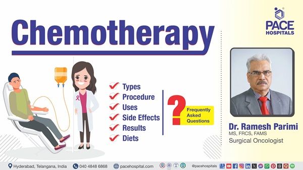
What is chemotherapy, and how is it administered?
Chemotherapy is a treatment that uses medications to kill or slow the growth of cancer cells. It can be administered intravenously (IV), orally (pills), through injections, or directly into affected areas like the spinal fluid. The treatment is given in cycles, allowing the body to recover between sessions.
What is targeted therapy, and how does it differ from chemotherapy?
Targeted therapy, also known as molecularly targeted therapy, is a cancer treatment that uses drugs or other substances designed to target specific proteins or molecules in cancer cells, thereby attacking them. Targeted therapy specifically attacks cancer cells by interfering with molecules involved in cancer growth, while chemotherapy affects all rapidly dividing cells, including healthy ones. Targeted therapy often has fewer side effects than chemotherapy and is usually based on mutations present in the cancer cells.
What is localized treatment, and how does it differ from targeted therapy?
Localized treatment focuses on a specific or limited body area, such as surgery or radiation therapy, to remove or destroy cancer cells in a particular region. Other examples include cryotherapy, laser therapy, and topical therapy.
On the other hand, targeted therapy is a systemic treatment that uses drugs designed to attack specific molecules or pathways involved in cancer growth, sparing normal cells. While localized treatment is helpful for tumours confined to one area, targeted therapy is effective against cancers that have spread or have specific genetic markers. Both approaches can be combined for better treatment outcomes.
When should I seek my oncologist appointment?
You will be referred to a medical oncologist if your healthcare professional suspects you have cancer or if you present with signs of cancer. You can consult a medical oncologist if you have any questions about cancer or experience any cancer-related symptoms such as a lump in any area, unexplained weight loss, fatigue and changes in skin colour.
Oncologists can perform diagnostic tests to confirm and understand the cancer and develop an effective treatment plan based on the cancer stage, type and extent of spread.
How to book an appointment in the Top Oncology Hospital in Hyderabad?
Anyone diagnosed with cancer and need a second opinion and guidance to start with cancer treatment or looking for an appointment in the best cancer hospital in the locations near Hitech City, Madhapur, Kondapur, Gachibowli, KPHB, or Kukatpally can visit the PACE Hospitals' Oncology Department Page and fill out the Appointment Form. They can also directly visit the hospital located near the Hi-Tech City metro station or call 04048486868 to book a hassle-free appointment with the Best Cancer Specialist in Hyderabad.
Types of Cancer Treated at PACE Hospitals
PACE Hospitals is ranked among the top 10 cancer hospitals in Hyderabad, Telangana, India, offering advanced care for a wide range of oncological diseases. Our team of expert oncologists and radiologists specializes in diagnosing and treating cancers of the digestive system, breast, reproductive organs, lungs, skin, kidneys, endocrine system, nervous system, and blood & lymphatic system.
We deliver patient-focused cancer care for conditions such as stomach cancer, esophageal cancer, colorectal cancer, cervical cancer, uterine cancer, ovarian cancer, breast cancer, lung cancer, leukaemia, myeloma, lymphoma, liver cancer, pancreatic cancer, gallbladder cancer, bladder cancer, prostate cancer, kidney cancer, oral cancer, and thyroid cancer using the latest technologies and compassionate, comprehensive treatment protocols.

Anal Cancer
Anal cancer is a type of cancer where the cancerous (malignant)cells form in the tissues of the anus. Anus is present at the end of the large intestine and below the rectum, where stools (solid waste) leave the body. Two ring-like muscles known as sphincter, which close and open the anal opening, let the stool pass out of the body. The skin around the outside of the anus is called the perianal area.
Anal cancer may develop in any part of the anus. Signs and symptoms of this cancer include a change in bowel habits, a lump near the anus, bleeding from the anus or rectum, and pain or pressure in the area around the anus. Most of these cancers are caused by Human papillomavirus (HPV) infection.
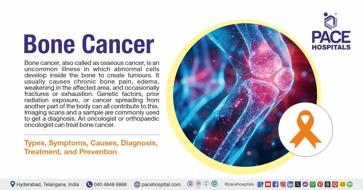
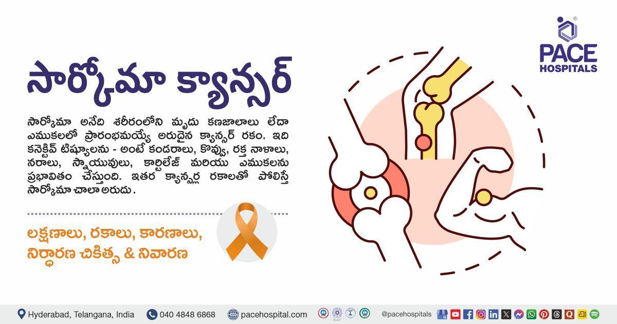
Bone Cancer
Bone cancer is an uncommon cancer that develops in the bones. It can affect any bone, but in most cases, it affects the long bones of the legs or upper arms. Common symptoms include persistent bone pain that worsens over time. Other symptoms are swelling and inflammation over a bone, leading to difficulty in movement if the cancer cells affect near a joint, a bone lump and a weak bone that fractures (breaks) more easily than normal bone. Common types of bone cancer include Osteosarcoma, Ewing sarcoma and chondrosarcoma.
Breast Cancer
A breast is formed by three main parts such as lobules (glands that make milk), ducts (that deliver milk to the nipples), and connective tissue (consists of fatty tissue and fibrous), which covers and maintains everything together.
Breast cancer is a severe condition in which cells in the breast grow uncontrollably. Cancer cells can grow, divide, and spread into nearby breast tissue, causing lumps or thickening. The type of breast cancer may vary depending on the location of the cells in the breast. Breast cancer cells begin inside the milk-producing lobules and the breast's milk ducts. If it is diagnosed earlier, it won't become life-threatening.
Brain Tumour
A brain tumour is considered the abnormal development of cells in the brain. Over 120 distinct types of tumours can emerge in the brain, and they can develop anywhere in the brain or skull and many other areas.
The brain contains different parts that operate different nervous system functions, and some tumours can spread into the spinal cord through the spinal fluid. Brain tumours can be either cancerous (malignant) or non-cancerous (benign). It's important to note that not all brain tumours are cancerous, but all brain cancers are tumour growths. Brain tumours are considered serious if they put pressure on or spread into healthy brain parts, but they rarely become cancerous. Examples of benign tumours include meningioma, vestibular schwannoma and pituitary adenoma.
Brain malignant growths tend to grow and penetrate the nearby, healthy brain structures. They can be life-threatening because of changes they cause to the essential structures of the brain. Examples of brain cancer include olfactory neuroblastoma, chondrosarcoma and medulloblastoma.
Bladder Cancer
Bladder cancer is a condition of the formation of cancer in the tissue of the urinary bladder. It starts with the development of cancerous cells that eventually form a tumour and spread to other parts of the body.
The bladder is a hollow organ that temporarily stores urine, which is located in the lower pelvis. A common type of urinary bladder cancer is urothelial or transitional cell carcinoma (TCC). The presence of blood in urine is the most common symptom, and others include a sudden urge to urinate, swelling of legs, a burning sensation while passing urine, unintentional weight loss and pelvic pain.
Colorectal Cancer
Colorectal cancer occurs in the tissues of the colon or rectum, which are called colorectal or colon or rectal cancer, depending on where they begin to grow. The colon is called the large intestine or large bowel, where the rectum is a passageway connecting the colon with the anus.
Colon and rectal cancer frequently group together because of common features. Colorectal cancer starts as a polyp in the rectum or colon. Over time, these polyps can turn into cancer. Diagnosing and removing them can prevent colorectal cancer. Early diagnosis is key in preventing cancer.
Chest Wall Cancer
The chest wall is the cavity (place) where the heart, lungs and liver are protected. Some tumours (growths) can originate from this place or spread elsewhere. This tumour may be cancerous or noncancerous; only 60% of tumours are cancerous. Any age group of people can get this cancer. However, older people have a high risk of developing cancerous tumours.
Signs and symptoms of this condition include a feeling of lump, fever and malaise, troubling movement, pain, swelling, tenderness, muscle atrophy, and sudden weight loss. The important note is that signs and symptoms are not visible until the tumour is in progressed condition, and they can depend on the tumour type. The exact cause of this condition still needs to be understood clearly.
Cervical Cancer
Cervical cancer is a type of cancer that begins in the cervical cells. Usually, this cancer develops slowly, and before the cancer begins in the cervix, the cells go through specific changes known as cervical dysplasia, where the abnormal cells start appearing in the cervix's tissue. If it is not removed or destroyed, it may cause cancerous cells to grow and spread in the cervix.
The most common cervical cancer cause is a long-lasting infection with human papillomavirus (HPV). People who start having sex before age 18 or within a year of the first period, have a weak immune system, cigarette smoking, use birth control pills, have sexually transmitted diseases (STDs) and have multiple sexual partners have an increased risk of getting this condition. Symptoms include painful sex, unusual vaginal bleeding and unusual vaginal discharge.
Cholangiocarcinoma (Bile Duct Cancer)
Cholangiocarcinoma, also called bile duct cancer, is an uncommon (rare), aggressive form of cancer that develops in the bile ducts, a network of tubes that transports bile to the small intestine from the liver. Jaundice, which is the yellowing of the skin and eyes, along with symptoms such as itching, weight loss, fever, abdominal pain, and light-coloured stools or dark urine, can indicate bile duct cancer. Treatment options usually involve a combination of chemotherapy or radiation therapy and surgery.
Endometrial Cancer
Endometrial cancer (uterus cancer) is the most common gynaecological cancer. It begins in the inner lining of the uterus called the endometrium. It is curable if it is diagnosed in the early stages.
Being obese and having metabolic syndrome may increase the risk of endometrial cancer. Signs and symptoms include painful sex, painful urination, abnormal vaginal bleeding, which is not related to the menstrual periods, post-menopausal bleeding and pain in the pelvis.
Fallopian Tube Cancer
Fallopian tube cancer is a similar type of ovarian cancer. It develops in the fallopian tubes, which connect the ovaries to the uterus. The exact cause of this condition is not known(idiopathic). Some possible risks are having a family history of breast or ovarian cancer.
Symptoms of fallopian tube cancer might include a swollen abdomen, a watery vaginal discharge that may present with blood, abdominal pain, vaginal bleeding not related to menstruation, and a lump or swelling in the lower abdomen.
Gallbladder Cancer
Gallbladder cancer is an uncommon (rare) form of cancer where cancer cells form in the tissues of the gallbladder. The gallbladder is an organ that is pear-shaped and lies under the liver in the abdomen, and it stores a fluid called bile (made by the liver) to digest fat.
This cancer begins in the inner layer of the gallbladder and multiplies and spreads to the outer layer of the gallbladder as it grows. Jaundice, fever, and pain are common symptoms of gallbladder cancer, which can be difficult to diagnose as it looks like symptoms of other medical illnesses.hat site visitors who are interested get more information. You can emphasize this text with bullets, italics or bold, and add links.
Head and Neck Cancer
Head and neck cancer is a group of cancers that can damage the various parts of the mouth, throat, and other areas in the head and neck. This condition can occur in the oral cavity, throat (pharynx), voice box (larynx), nasal cavity, and salivary glands. They can also begin in the muscles, sinuses, or nerves in the head and neck.
It can be caused by human papillomavirus infections, especially HPV 16, radiation exposure, Epstein Barr virus infection, underlying disorders and use of alcohol, tobacco and paan (betel quid). The most common symptom of this cancer includes persistent sore throat, and other symptoms include a lump in the throat, mouth or neck, headache, neck pain, hoarseness, and red or white patches on gums, tongue or inside the mouth.
Hypopharynx Cancer
Hypopharynx cancer is the type of cancer that is characterised by the development of cancerous (malignant) cells in the tissues of the hypopharynx. The hypopharynx is a (bottom part of the pharynx) 5-inch-long part of the throat that prolongs behind the nose, down to the top of the trachea and oesophagus.
Smoking and chewing tobacco products, heavy alcohol consumption, and having Plummer-Vinson syndrome can increase the risk of developing hypopharyngeal cancer. Signs and symptoms of this condition include ear pain and sore throat. Other symptoms include difficulty or painful swallowing, a lump in the neck and changes in the voice.
Hodgkin Lymphoma
Hodgkin lymphoma is a rare type of cancer originating in the lymphatic system, which is a network of glands and vessels where cancer can divide and spread throughout the body. The lymphatic system is a vital part of the immune system. It is responsible for the movement of lymph (a fluid containing infection-fighting white blood cells called lymphocytes) through lymphatic vessels, making it an essential part of the immune system.
In Hodgkin lymphoma, abnormal multiplication of B-lymphocytes occurs and is collected in specific parts such as lymph nodes of the lymphatic system. The affected lymphocytes can lose the infection-fighting property, making them more vulnerable to infection. The main symptom of Hodgkin lymphoma is a painless swelling in a lymph node, commonly in the neck, armpit, or groin.
Hepatobiliary Cancers
Hepatobiliary cancers are a varied group of malignant cancers that affect the liver, gallbladder, and bile ducts. The most common types include hepatocellular carcinoma (HCC), which originates in liver cells, and cholangiocarcinoma, which arises in the bile ducts. Gallbladder cancer is another form of hepatobiliary cancer. These cancers are often linked to chronic liver conditions, including hepatitis B or C, cirrhosis, and fatty liver disease.
Symptoms may include jaundice, abdominal pain, and unexplained weight loss. Early diagnosis is challenging, and treatment options may include surgery, chemotherapy, radiation, intra-arterial therapies or liver transplantation
Kidney Cancer
Kidney cancer, also known as renal cancer, is characterised by the development of cancer cells in the tissues of the kidneys. Kidneys are a pair of bean-shaped organs attached to the upper back side wall of the abdomen, protected by a lower rib cage. They act as excretory organs by removing the excess amounts of salt, water and waste products from the body.
Some conditions increase the risk of kidney cancer, such as smoking, a family history of kidney cancer, obesity, high blood pressure and long-term dialysis for chronic kidney disease.
Symptoms include blood in urine, a lump or swelling in the back, tiredness, sweating, loss of appetite and weight loss.
Kaposi Sarcoma
Kaposi sarcoma is a cancer that grows from the cells lining blood or lymph vessels. It is characterized by the growth of purple-coloured lesions made of cancerous cells, white and red blood cells, and new blood vessels. These lesions may arise in multiple locations of the body and contain human herpesvirus (HHV-8), called Kaposi sarcoma herpesvirus (KSHV), but in most cases, people with HHV-8 do not get Kaposi sarcoma. Still, they can have a risk of developing Kaposi sarcoma if their immune system is weakened by medications that are given after an organ transplant or certain other medical conditions such as human immunodeficiency virus (HIV).
Lung Cancer
Cancer is referred to as the unlimited growth of cells. If it occurs in the lungs (bronchi or alveoli), it is called lung cancer, and this type of growth is usually seen in bronchi or alveoli. It starts in the lungs and may spread to the other organs in the body or lymph nodes, and cancer from other organs may also come to the lungs.
Mainly lung cancers are divided into two types: non-small cell (more common) and small cell lung cancer; they grow in different manners. So, lung cancer treatment and symptoms depend on the spreading and stages. symptoms include coughing up phlegm, wheezing, weakness, loss of appetite and weight loss, chest pain and recurrent respiratory infections.
Liver Cancer
Primary Liver cancer is a condition characterised by the formation(growth) of cancerous cells in the tissues of the liver. Secondary liver cancer is a separate condition where the cancer initially develops (grows) in other parts and spreads to the liver.
The liver is one of the largest organs in the body, has two lobes and is located at the upper right side of the abdomen inside the rib cage. It helps in digesting the food and removal of toxins. Hepatocellular carcinoma is the main type of liver cancer. It is the third leading cause of death (cancer-related) worldwide, and another main type is called cholangiocarcinoma (bile duct cancer). Signs and symptoms of this cancer include a swollen abdomen and easy bruising, loss of appetite, a hard lump on the right side below the ribcage, weight loss and fever.
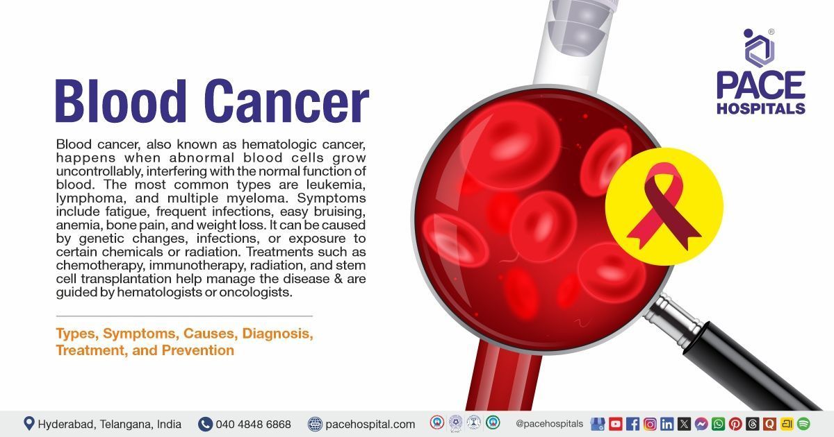
Leukaemia
Leukaemia is also called blood cancer, caused by increased WBC (white blood cells) in the body. Cancer cells that start to grow(form) in the blood-forming tissue of the bone marrow are called leukaemia’s. These do not form as solid tumours, but large amounts of abnormal white blood cells can accumulate in blood and bone marrow, crowding out of the normal cells (red blood cells), resulting in less oxygen to tissues.
There are four types of leukaemia cancers:
- Acute lymphocytic leukaemia,
- Chronic lymphocytic leukaemia,
- Acute myeloid leukaemia and Chronic myeloid leukaemia.
Symptoms include weakness, easy bruising or bleeding, vomiting, weight loss and pain in bones or joints.
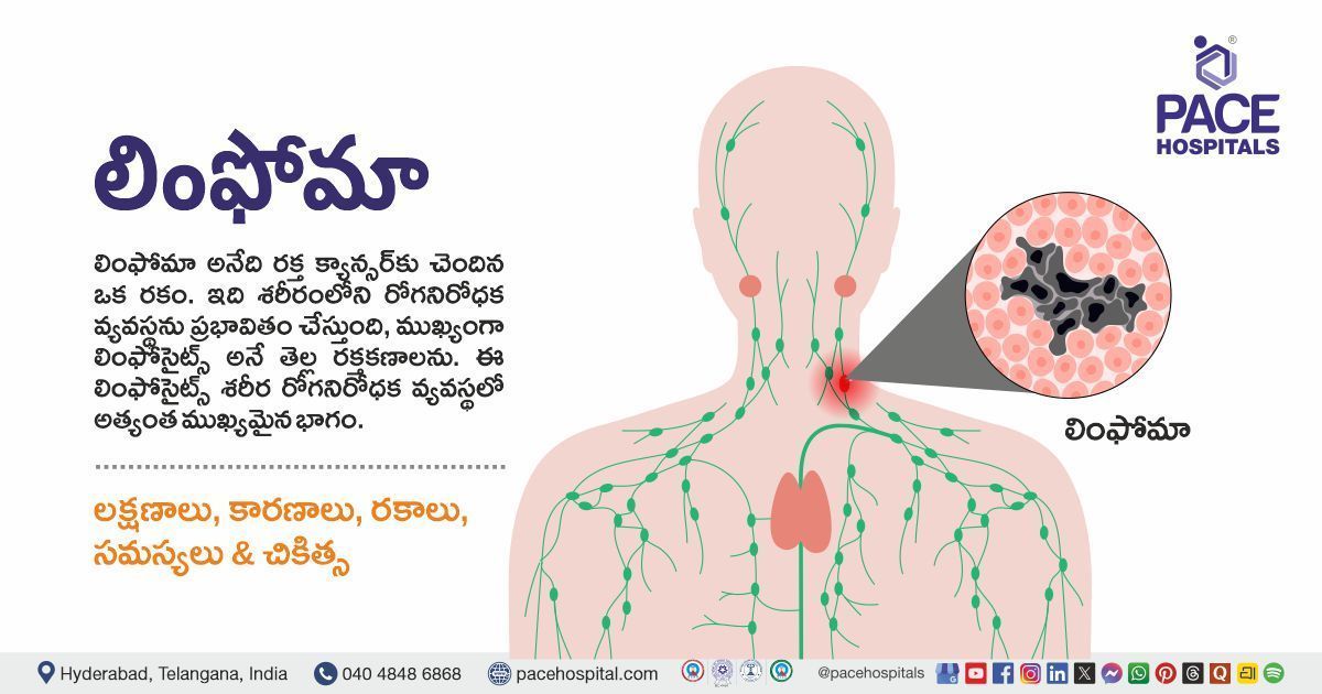
Lymphoma
Lymphoma is a cancer characterised by the formation of cancer cells in the lymphatic system. The lymphatic system compromises the spleen, thymus, bone marrow, and lymph nodes, removes excess fluids from the body and produces immune cells. There are two types of lymphoma, including Hodgkin lymphoma and non-Hodgkin lymphoma.
Symptoms include fever, cough, fatigue, weight loss, and swollen glands (lymph nodes), often in the armpit, neck or groin, which are painless. Lymphoma and leukaemia are both different cancers. Each develops in a different type of cell as leukaemia starts in blood-forming cells in the bone marrow, and lymphoma grows in infection-fighting cells of the lymphatic system.
Lip Cancer
Lip and oral cancers usually result from abnormal growth of surface layer squamous cells in the mouth and lips. They are characterised by the formation of cancer (malignant) cells in the lips or mouth and oral cavity.
Too much exposure to UV light or sunlight, heavy alcohol consumption, tobacco use and being male can raise the chances of developing lip cancer. Symptoms include bleeding, thickening of the lip, lesion, blister, lump or sore in the lip, discoloured patch on the lip and lip pain or numbness.
Laryngeal Cancer
Laryngeal cancer is a condition where cancerous (malignant) cells develop in the tissues of the larynx.
Having too much alcohol, usage of tobacco products and exposure to certain chemicals such as coal dust and asbestos can increase the risk of laryngeal cancer.
Signs and symptoms of this condition include ear pain, sore throat or cough, pain or difficulty while swallowing, a lump in the throat or neck and change or hoarseness in the voice. Treatment options include radiotherapy, chemotherapy, surgery and anti-cancer medications.
Melanoma
Melanoma is a type of cancer where cancer (malignant) cells develop in the melanocytes, which are the cells that give colour to the skin. Human skin is the largest organ in the body, and it gives protection against sunlight, heat, injury and infection. It also helps to control the body's temperature and stores fat, water and vitamin D. It comprises several layers. Skin cancer usually starts in the uppermost layer, the epidermis.
Signs include a change in coloured (pigmented) skin, asymmetrical mole that itches oozes, bleeds and has irregular edges. Usually, normal moles are in one colour, but often melanomas have a mix of two colours. Treatment options include surgery, radiotherapy, medications and chemotherapy.
Mesothelioma
Mesothelioma is a type of cancer that forms in the tissue layers covering the outer surface of some body organs called mesothelium and is commonly associated with asbestos exposure. Primarily affecting the lining of the lungs (pleural mesothelioma), this cancer can also impact other organs such as the stomach, heart, or testicles. It is more commonly diagnosed in men who are over the age of 75.
Symptoms depend on where cancer attacked, such as lungs (chest pain, a persistent cough, shortness of breath, clubbed fingertips) and stomach include (feeling sick, tummy pain, unexplained weight loss, diarrhoea or constipation).
Multiple Myeloma
It is also known as myeloma, a type of bone marrow cancer that attacks the bone marrow. Bone marrow is the soft tissue with bones inside, consisting of red blood cells (RBC), white blood cells (WBC). Cells which are primarily located in the bone marrow, make an abnormal antibody (protein) called monoclonal immunoglobin, monoclonal protein (M-protein).
Symptoms of this cancer include:
- Bruising and unusual bleeding.
- Blurred vision.
- Repeated infections.
- Persistent bone pain (usually in the hips or ribs).
- Anaemia.
- Weak bones that fracture easily.
Nasopharyngeal Cancer
Nasopharyngeal cancer is a cancer in which cancerous (malignant) cells form in the nasopharynx. The pharynx is a 5-inch-long hollow tube that begins from behind the nose and ends where the windpipe (trachea) and oesophagus begin. The nasopharynx (the upper part of the pharynx) presents behind the nose.
Nasopharyngeal cancer commonly develops in the squamous cells that line the nasopharynx. Having Asian or Chinese ancestry (Ethnic background), exposure to the Epstein-Barr virus, and alcohol consumption can increase the risk of nasopharyngeal cancer. Signs and symptoms of nasopharyngeal cancer include a sore throat, trouble hearing, pain or ringing in the ear, headaches, nosebleeds and a lump in the nose or neck.
Non-Hodgkin Lymphoma
Non-Hodgkin lymphoma is a disease characterised by the formation of malignant (cancer) cells in the lymph system. Non-Hodgkin lymphoma can have variable levels of aggressiveness ranging from slow-growing to fast-growing. Some factors, such as being male, having older age, and having a weakened immune system, can raise the risk of non-Hodgkin lymphoma.
Signs and symptoms of this condition include drenching night sweats, fever, weight loss, fatigue and swollen lymph nodes. Tests that examine the lymph system and different body parts are used to diagnose non-Hodgkin lymphoma. The treatment and prognosis depend on the patient's age, signs and symptoms, stage and type of cancer.
Oral Cancer
Oral cancer includes the development of cancer cells in the mouth and back of the throat. It can occur on the tongue, under the tongue, at the base of the tongue and on the tissue lining the mouth. Most cancers in the mouth are associated with alcohol consumption, smoking (tobacco use) or both and human papillomavirus (HPV) -(most common throat cancer) and other causes include age (most often occurs in over the age group people of 40), sun exposure, poor nutrition.
Common symptoms the affected person shows include a white or red patch in the mouth, a lump in the neck, difficulty chewing, swallowing or speaking, pain or bleeding in the mouth, earache, numbness in the tongue or other mouth areas, etc. This cancer can spread easily and quickly, so early identification is essential for effective treatment.
Oesophageal Cancer
Oesophageal cancer is a type of cancer characterised by the development of cancerous (malignant cells) in the tissues of the oesophagus. The oesophagus is the muscular hollow tube that helps move liquid and food from the throat to the stomach. Oesophageal cancer starts on the lining of the oesophagus; as it grows, it will spread outward through another layer.
Smoking, older age, Barrett oesophagus and heavy alcohol consumption can raise the risk of this cancer.
Signs and symptoms include weight loss, difficulty or painful swallowing, hoarseness and heartburn and a lump under the skin.
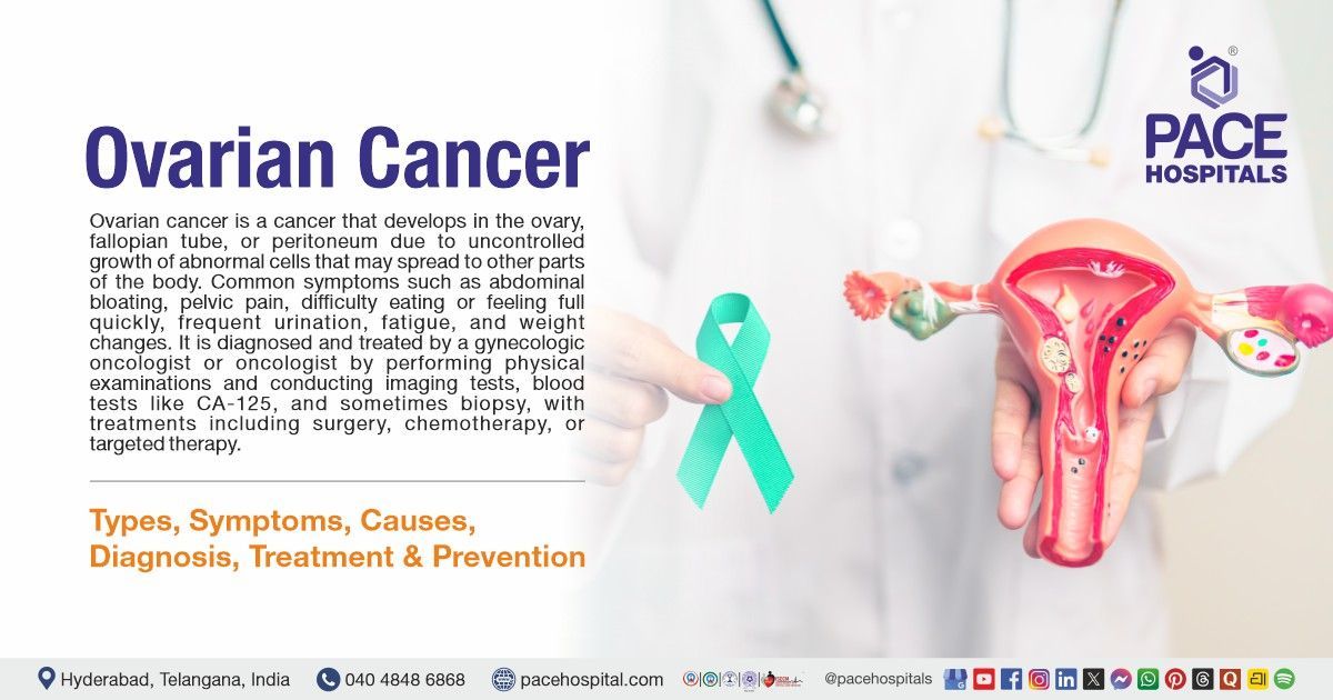
Ovarian Cancer
Ovarian cancer is a condition where cells in the ovary grow abnormally and develop into a tumour. If not detected early, the cells can spread to surrounding tissues and other areas of the body. It commonly affects women who are 50 or older.
Symptoms include pelvic pain or pressure, bloating, vaginal bleeding or discharge, difficulty eating, urgency to urinate, and abdominal or back pain. The exact cause is not understood completely, but studies have shown that having a family history of ovarian or breast cancer, older age, obesity, a history of breast cancer, and undergoing hormonal replacement therapy can increase the risk.
Penile Cancer
Penile cancer is an uncommon cancer where abnormal cell develops uncontrolled in the penis of the male. It can grow anywhere in the penis, but it is commonly seen under the foreskin of the penis. Penis is an essential part of the male reproductive system.
The exact cause is idiopathic (unknown), but having human papillomavirus and smoking can increase the risk of developing penile cancer. It is commonly seen in men over 50. Signs and symptoms include a growth or sore on the penis, a foul smelly discharge, a rash on the penis, and a change in the skin colour of the penis.
Pancreatic Cancer
Pancreatic cancer is a medical condition where cancerous (malignant) cells form in the pancreas. The pancreas is a six-inch-long gland that is pear-shaped and lies between the stomach and the spine.
It breaks down food and controls blood sugar levels by making certain hormones.
Smoking, being overweight, having a family history of pancreatic cancer, and having a history of diabetes or chronic pancreatitis can increase the risk of developing pancreatic cancer. Signs and symptoms include pain, jaundice and weight loss, but it can be challenging to diagnose early and often requires medical tests and procedures.
Prostate Cancer
Prostate cancer is a condition characterised by the formation(growth) of cancerous cells in the tissues of the prostate, which is part of the male reproductive system, including the penis, prostate, seminal vesicles and testicles.
The prostate is present below the bladder and in front of the rectum, producing semen. Its size changes as a man ages, and it is more common in older men. The common signs of an affected person are frequent urination or weak urine flow. Other signs and symptoms include:
- Trouble starting the urination.
- Difficulty emptying the bladder fully.
- Weak or interrupted urine flow.
If this cancer is identified in an advanced stage, symptoms include pain in the back, hips or pelvis, tiredness, shortness of breath or pale skin caused by anaemia. So early identification plays an essential role in treating it.
Pharyngeal Cancer
It is a type of cancer in which cancerous (malignant) cells form in the pharynx. The pharynx is a 5-inch-long hollow tube that begins from behind the nose and ends where the windpipe (trachea) and oesophagus begin.
The pharynx has three parts:
- Nasopharynx: upper part
- Oropharynx: middle part
- Laryngopharynx (hypopharynx): bottom part
Cancer in the oropharynx is called oropharyngeal cancer. The oropharynx is the region of the throat (pharynx) that starts from the back of the mouth. The oropharynx includes the soft palate, tonsils, side and back walls of the throat, and back one-third of the tongue.
Human papillomavirus (HPV) infection, alcohol consumption, chewing betel quid and smoking can increase the risk of oropharyngeal cancer. Signs and symptoms of this cancer include a sore throat, difficulty in moving the tongue, trouble swallowing, a lump in the neck and ear pain.
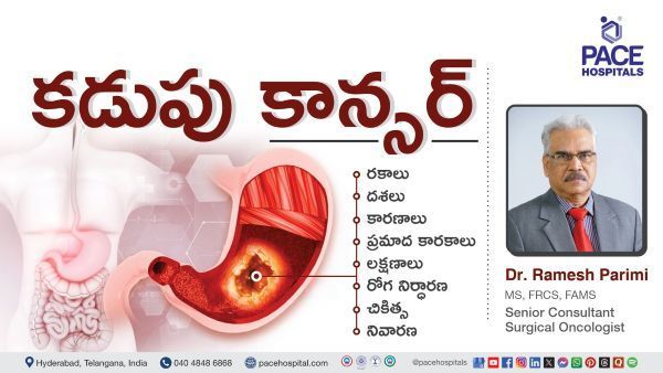
Stomach Cancer
Stomach (gastric) cancer is an uncommon form of cancer that develops in the lining of the stomach. The stomach plays a crucial role in digesting food and is part of the digestive tract, a long, twisting tube of hollow, muscular organs that run from the mouth to the anus.
Initially, these cancer symptoms are vague, and it is easy to mistake them for other health conditions such as heartburn, persistent indigestion, persistent stomach pain, and feeling full. Other advanced cancer symptoms include loss of appetite, blood in stools and weight loss.
Salivary Gland Tumours
Salivary gland tumours are characterised by the growth of abnormal cells in the salivary gland or in the ducts(tubes) that drain the glands. Salivary glands are the organs present on each side of the face, which are helpful in making saliva (spit), the lubricating fluid found in the throat and mouth, antibodies, and it has digestive enzymes.
Salivary gland tumours can be cancerous (benign) or non-cancerous(malignant). Most salivary gland tumours are non-cancerous(benign), but some are cancerous(malignant). If left untreated, benign tumours can become cancerous over time. Salivary gland cancer is the growth of cancer cells in the salivary glands, where the signs include a lump or trouble swallowing. Usually, the person with this cancer cannot show any signs and symptoms, but it may be found during routine tests or physical or dental examinations.
Squamous Cell Carcinoma
This type of cancer develops in squamous cells, which are thin, flat cells lining various body parts, including skin, respiratory tract, and digestive tract. It is commonly caused by prolonged sun exposure, smoking, and exposure to carcinogens. SCC can appear as a red, scaly patch, a sore that doesn't heal, or a lump. If this cancer is left untreated, it can spread to nearby tissues and organs. Treatment options for this cancer include surgical removal, immunotherapy, radiation therapy, and chemotherapy, with early detection offering better outcomes.
Thyroid Cancer
Thyroid cancer is a type of cancer where the cancer cells develop in the tissues of the thyroid gland. The thyroid gland is butterfly-shaped, an essential organ of the endocrine system, present at the front side of the neck just above where collarbones meet. It makes the hormones which can control the metabolism.
A lump or nodule can appear in a routine medical exam. A thyroid nodule (solid or fluid-filled) is an abnormal development of cells in the thyroid. If a healthcare professional finds nodules, they may conduct an aspiration biopsy to check for signs of cancer.
Thymoma and Thymic Cancer
Thymomas and thymic cancers are rare malignant tumours which form in the thymus cells. Thymomas have slower growth and rarely spread over the thymus; thymus carcinoma has faster growth and often spreads to the other body parts, and it is not easy to treat.
The exact cause of thymoma and thymic cancer is not understood fully, and research is ongoing to find any link between viruses, heredity and other cancers. Most people don't show any noticeable symptoms in the early stages, but they show the signs when the tumour affects the organs in the chest area. Common symptoms of these cancers include chest pain, hoarse voice, trouble breathing, persistent cough and lack of appetite.
Testicular Cancer
The male reproductive system comprises a penis, scrotum with testicles and prostate. Testicles are a pair of tiny egg-shaped glands located in the scrotum below either side of the penis that aid in the formation and maturation of sperm.
Cancer that develops in the testicle, called testicular cancer, occurs when the cells in the testicle develop to form a tumour. This is considered one of the less common cancers and is most common in men between the ages of 15 and 49, and 90% of cases originate in the germ cells producing sperm.
Signs and symptoms include a lump in the testicle (painless), swelling of the testicle, dull pain in the lower abdomen or groin, breast tenderness or growth, and the sudden buildup of fluid in the scrotum.
Tracheal Cancer
It is an uncommon kind of cancer that arises in the trachea, the windpipe that joins the throat and lungs. Adenoid cystic carcinoma and squamous cell carcinoma are the two most prevalent varieties. Symptoms often include persistent cough, difficulty breathing, wheezing, and coughing blood. Smoking and prolonged exposure to environmental toxins are major risk factors.
The treatment plan depends on the size, stage and tumor location, the patient's overall health, and other factors. Tracheal cancer treatments include surgery, radiation therapy, chemotherapy, and bronchoscopic treatments.
Vaginal Cancer
Vaginal cancer is uncommon cancer where malignant cells form anywhere in the vagina (the tube that connects the vulva and the cervix). It is often caused by infections such as human papillomavirus (HPV).
Initially, vaginal cancers do not show any signs or symptoms. Still, some people present with symptoms such as vaginal discharge, blood in the urine or stool, constipated feeling, pain in the pelvis, painful urination and painful sex.
Vulvar Cancer
Vulvar cancer is an uncommon cancer characterised by the development of cancerous (malignant) cells around the tissues of the vulva located outside the body. It most commonly affects the outer lips of the vulva. Having HPV infection or vulvar dysplasia and other risk factors include:
- Having multiple sexual partners.
- Having first sex at a young age.
- Having a history of genital warts and being older can increase the chances of getting vulvar cancer.
Signs and symptoms include a lump or growth on the vulva that looks like an ulcer, itching around the vulvar area, pain in the vulvar area, and bleeding.
What Our Patients Say – Google Reviews
Advanced Cancer Diagnostics & Procedures
At PACE Hospitals, our skilled and experienced cancer specialists are dedicated to delivering the
best cancer treatment in Hyderabad, Telangana, India. We utilize advanced diagnostic tools like PET-CT, MRI, and counselling for early and precise detection—helping determine cancer grade and stage accurately. Our team of medical and surgical oncologists specializes in minimally invasive and robotic cancer surgeries, chemotherapy, immunotherapy, targeted therapy, and precision medicine, ensuring personalized, multi-modal treatment strategies for optimal outcomes.

Clinical Laboratory Tests for Cancer
Blood Tests
Blood tests play a vital role in diagnosing and treating cancers. It can help give important information on the overall health and function of the kidneys and liver. It can identify cancers by identifying certain chemicals and proteins in blood. But blood tests alone cannot identify cancer, so additional tests must be recommended to detect cancers.
Blood Chemistry Test
It measures the amounts of specific substances released into the blood by tissues and organs. These substances include fats, sugars, electrolytes, metabolites, proteins and enzymes. Blood chemistry tests provide crucial information on the liver, kidneys, and other organs' functions. Abnormal levels of substances in the blood indicate disease or cancer or treatment side effects.
Complete Blood Count (CBC)
Complete blood count measures the number or amounts of white blood cells (WBC), red blood cells (RBC), haemoglobin (a protein that carries oxygen in the blood) in the blood. It also measures the haematocrit value (amount of blood made up of red blood cells) and the size of red blood cells. It is often part of routine health check-ups and can help diagnose some cancers, especially leukaemia’s, monitors blood count during the treatment of cancer.
Cytogenetic Analysis
Cytogenic analysis examines the chromosome changes in blood, tissue, bone marrow or amniotic fluid samples. Chromosome Changes may contain missing, broken, rearranged or extra chromosomes. Changes in specific chromosomes may reveal signs of certain types of cancer.
Cell Search Circulating Tumour Cell (CTC) Test
This kind of blood diagnostic test is used to detect the circulating tumour cells (the cells from a malignant (cancerous) tumour that enter the blood), which is helpful in identifying prostate, breast and colorectal cancers.
Immunophenotyping
This test detects cells using antibodies that bind to specific antigens or markers on the cell surface. It is commonly performed on blood or bone marrow samples. Sometimes, it may be performed on other body tissue samples or body fluids. It helps identify, stage and monitor blood disorders and blood cancers such as lymphomas, leukaemia’s, myeloproliferative disorders and myelodysplastic syndromes.
Liquid Biopsy
Liquid Biopsy is a test performed on a blood sample to look for cancer cells or fragments of DNA from a tumour that may have been released into the bloodstream. This procedure may help identify the cancer at an early stage and help plan the treatment, detect whether the given therapy is working, and monitor the recurrence of cancer.
Sputum Cytology
Sputum cytology is a simple, non-invasive, and safe diagnostic process that identifies and counts the cell types and its morphology to assess the normal and diseased characteristics of the mucosa. This method aids in the identification of abnormal cells, inflammatory cells (lymphocytes, neutrophils, eosinophils, mast cells), bacteria, and hyphae/spores of fungi in the sputum. It helps in diagnosing lung cancer.
Tumour Marker Tests
Tumour marker tests are diagnostic examinations that evaluate the cancerous and non-cancerous substances produced by cells in response to cancer. Tumour markers are substances highly produced by cells in the body when benign conditions or cancer is present. These substances are proteins (mostly), but changes in DNA are sometimes used as tumour markers. The substances may be identified in urine, blood, stool or tumour tissue. Most of the tumour markers were made by both normal and cancer cells. It is important to note that cancer cells tend to generate higher levels of tumour markers than normal cells. They aid in diagnosing cancer, deciding the therapy (treatment), monitoring treatment progress (checking whether treatment is working), evaluating effectiveness (finding how well the treatment works on a person), and looking for signs of cancer that have reoccurred.
Urinalysis
Urinalysis is a diagnostical test that evaluates urine's characteristics, including its colour and contents. This test can identify the abnormal levels of sugars, protein, red blood cells and white blood cells. By studying these components, urinalysis can provide information about health and help diagnose bladder, kidney, and urothelial cancers.
Urine Cytology
Urine cytology discovers illness by looking for abnormal cells shed into the urinary tract's urine. Urine cytology enables analysis of bladder, kidney, and more infrequent urothelial cancers. After cancer treatment, it is used to look for signs that cancer has returned.
CA-125 Test
CA 125 test is called the cancer-antigen 125 test, which checks for specific proteins in the blood. This blood test measures the total amount of protein called CA-125 (cancer antigen 125) in the body. It is a type of blood biomarker representing a disease or condition. It is often used as part of a complete approach to monitor ovarian and other cancers. It is a screening examination for people at high risk of developing ovarian cancer. It cannot detect cancer, but it can monitor certain types of cancer during and after cancer treatment.
Prostate-Specific Antigen Test
A prostate-specific antigen test, also called a PSA test, is a protein produced by normal (healthy) and malignant cells of the prostate gland. The level of PSA is often increased in the blood of prostate cancer patients. Other conditions also increase the levels of PSA, such as prostatitis and BPH (benign prostatic hyperplasia). The PSA test estimates the amounts of PSA in the bloodstream. A healthcare provider must send a blood sample to the lab for further analysis during the test. Men with symptoms linked with prostate cancer may have a prostate-specific antigen test and a digital rectal exam (DRE).
Imaging Tests for Cancer
Computed Tomography (CT Scan)
A CT scan utilizes an X-ray machine linked to a computer system to take a series of images of organs from different angles, which helps create detailed 3D pictures of the inside of the body. Before the scan, dye or contrast material can be used, or gives a needle into a vein, gives dye to swallow, which helps produce the images by highlighting specific areas in the body. The CT machine moves around the patient during the CT scan by creating the images. It will help diagnose colorectal cancer and lung cancer.
Magnetic Resonance Imaging (MRI Scan)
An MRI is an imaging instrument that uses powerful radio waves and magnets to take images of the body in slices. These slices are merged to create detailed cross-sectional pictures of the inside of the body to distinguish between normal and diseased tissue, which can show the places where the tumours are located and also reveal the metastases. It is often used for imaging of the connective tissue, brain, muscle, spine and inside of bones. During the process, the patient has to lie on a table and is pushed into a long chamber, where the MRI makes loud thumping noises and rhythmic beats. A special dye (contrast agent) was injected into a vein, which shows the pictures more brightly.
Nuclear Scan
A nuclear scan is a diagnostic imaging technique that uses some radiation to capture pictures of the body's bones, tissues, and organs. It is also called a radionuclide scan. A machine called a scanner measures the radioactivity in the body. It creates images of specific organs or bones on a computer screen or film. During this scan, a small amount of radioactive material (tracer) is injected into the individual, which flows through the bloodstream, reaches the tumour and sticks to it, where it "light up" to see through a special scanner. These imaging tests diagnose and treat specific cancers such as breast, brain, kidney, thyroid, liver, bladder, lung and bone cancers.
Bone Scan
Bone scan, also known as scintigraphy, is a nuclear test used to diagnose bone abnormalities or damage and determine the severity of diseases. It shows where the cancer starts in bones or spreads to bones. This test helps to measure the progress of treatments for bone cancer and helps to detect the cancer in an earlier stage than regular x-rays. Specific kinds of cancer are known to spread to bones, including lung, breast, prostate, and kidney.
Positron Emission Tomography (PET Scan)
A PET scan is a nuclear imaging scan that gives detailed 3D images of areas inside the body using radioactive sugar. Because cancer cells often consume more glucose than normal cells, these images can be used to diagnose cancer in the individual. Before the process, a radioactive glucose tracer is injected into the patient, which is helpful in identifying the cancers in the body.
PET/CT Scan
Healthcare professionals commonly combine PET with CT scans to accurately diagnose tumours. The PET scan provides information on areas of elevated cell activity, while the CT scan shows more precise images of these areas.
X-Rays
X-rays (radiographs) are a non-invasive diagnostical instrument that uses low radiation to create images of the body's internal bones, tissues and organs onto the film. X-rays use invisible electromagnetic energy beams to produce pictures of internal organs of the body. A healthcare provider will put the patient in a particular position and direct the X-ray beam to a specific part of the body. It is used to help identify and stage the cancer.
Ultrasound
An ultrasound, also known as ultrasonography or sonography, utilizes high-energy sound waves to produce images of the internal organs of the body. When sound waves contact the body's organs, they are sent back by bouncing off them. A tool called a transducer turns the sound waves into images. An ultrasound is helpful in finding the tumour to healthcare professionals by showing the exact location of the tumour in the patient's body. The healthcare professional can see tumours by examining the echoes produced by sound waves as they pass through the body's tissues.
Mammography (Mammogram)
Mammography is an X-ray imaging process that looks for cancer in breast tissue. It is used as both a screening mammography and diagnostic mammography. Screening mammography is a routine examination performed every one or two years to look for breast cancer with no signs or symptoms. Screening mammography aims to identify the cancer early, which is easy to treat. A diagnostic mammogram is performed to investigate symptoms such as a lump in the breast or chest and other signs or when the health care professional wants more information about the patient's breast health.
MUGA Scan
Cardiotoxicity is a side effect of some cancer treatments, which can cause heart problems. MUGA is a nuclear imaging test also called equilibrium radionuclide angiocardiography (ERNA) or radionuclide ventriculography (RNVG). MUGA scan examines the function of the heart. It may used to investigate heart function before, during and after the chemotherapy. It measures the amount of blood pumped out of the heart (ejection fraction). If it shows abnormal values, the healthcare professional may change the chemotherapy type. It can help to choose the best treatment for individuals with heart issues. Specific cancer treatments can cause chronic damage to the heart. A healthcare professional may recommend a MUGA scan or other scans to look at the heart if someone has had:
- Radiation therapy for chest
- Certain types of chemotherapy and immunotherapy
Lymphangiogram
A lymphangiogram is a medical imaging examination that uses X-ray or MRI techniques to create images of the lymphatic system and identifies abnormalities or cancerous (malignant) cells in the lymphatic system and structures. It involves an injection of dye into the lymph system to visualize and create pictures of lymphatic structures.
Thyroid Scan
A thyroid scan is a type of nuclear imaging examination that is a painless diagnostic technique that utilises radioactive substances called radiopharmaceuticals or radiotracers. Radiotracers contain tiny portions of radioactive material. They can collect in tumours and areas of inflammation and attach to specific proteins in the body. The patient receives a radiotracer through injection, swallow or inhalation. It accumulated in the regions being examined; a special camera identifies the gamma rays emitted from the radiotracer and creates and produces images and molecular information on the computer. This scan is used to detect cancers of the thyroid.
Gallium Scan
A gallium scan is a nuclear imaging scan that can discover inflammation, infection, or cancer in the body. It creates pictures of the body by using radioactive materials. These materials make radiation where special machines can detect the energy. A radiologist injects a small amount of radioactive material into a patient's blood. The gallium collects (settles)in regions of the body with infection or inflammation. A particular camera locates the gallium and takes pictures. A gallium scan can help detect cancer (Hodgkin lymphoma), inflammatory conditions (pulmonary fibrosis or sarcoidosis) and infection (abscess, osteomyelitis).
Endoscopic & Interventional Diagnostic Tests
Pap Test
It is also called a pap smear test, a screening procedure to assess cervical cancer. It identifies the presence of precancerous or cancerous cells. It can determine the changes in the cells on the opening of the uterus called the cervix. This test is usually performed by inserting a metal or plastic tool named a speculum into the vagina to open and widen the vagina walls. Health care professional uses a swab to take a sample piece of cells from the cervix to send it for lab evaluation.
Biopsy
In most cases, healthcare professionals must perform a biopsy to confirm the cancer. A biopsy is a minor procedure of removal of a sample of abnormal tissue, which is examined under the microscope and other examinations on the cells in the sample. Biopsy samples may be removed in several ways.
With a needle: Healthcare professional uses a needle to withdraw fluid or tissue. This method is performed to take some breast, prostate and liver biopsies.
With an endoscopy: Healthcare professionals insert a thin, lighted tube known as an endoscope into a mouth or anus (opening of the body) and remove some or complete abnormal tissue through the endoscope.
Fine-Needle Aspiration
FNA (Fine-needle aspiration) is a procedure that healthcare professionals use to get a cell sample from an abnormal area or suspicious lump. It may also be performed during a bronchoscopy or endoscopy. It uses a thin needle and syringe to pull(take) out the abnormal cells, tissue and fluids. It will identify the masses (tumours)in the breast, skin, lymph nodes and thyroid.
Colonoscopy
A colonoscopy is a medical diagnostic procedure that allows the healthcare professional to examine the large intestine (colon), which helps identify potential problems such as polyps or colorectal cancer. Polyps are small masses that are non-cancerous, but they become cancerous. This test uses a small instrument called a colonoscope, which will pass into the rectum tube and then into the colon and remove a tissue sample for further evaluation.
Bronchoscopy
It is a procedure that looks inside the lung airways and is also used to find the causative factor of a lung problem. It can recognise signs of infection, tumours, bleeding, excess mucus in airways and lung blockages. A healthcare professional will slide a small tube attached to a camera at the end of the airways. By looking into the camera, they can see the passages inside, such as blood, mucus or tumours. A medicine to sleep or a drug should be given to numb air passages before performing this procedure. If an imaging study found any abnormal cells in a patient's lung, a piece of the lung growth may be taken to detect a cancerous tumour or another condition.
Mediastinoscopy
A mediastinoscopy is a surgical procedure involving a mediastinoscope to examine the mediastinum, the space between the lung lobes behind the breastbone. The mediastinoscope contains a small camera to provide a visual of the area. However, healthcare professionals must create a small hole in the chest to insert the device. This procedure is typically performed to identify signs of various cancers and to diagnose conditions like inflammation, sarcoidosis, thymoma, and cancer affecting the bronchi or other structures in the mediastinum. It is often performed to biopsy or remove the lymph nodes in the space between the lungs to identify cancer or to stage lung cancer, thymoma, oesophagus cancer or lymphoma.
Pleural Biopsy
The lungs are surrounded by two layers of membranes, known as the pleural cavity. Sometimes, certain illnesses can lead to fluid accumulation in the area between the lungs and the pleura. If this occurs, a Healthcare professional may suggest a test to check for any fluid buildup. This test involves using a specialized biopsy needle to extract a small piece of pleura. Its purpose is to diagnose infections, cancers, or other diseases.
Thoracoscopy
A thoracoscopy is a medical procedure that utilizes a thin, flexible tube with a light and attached camera at the end to examine the cavity within the chest and the surface of the lungs. It is performed to identify the underlying causes of lung problems, such as coughing up blood and difficulty breathing, and to obtain tissue samples (biopsy) from abnormal lung tissue, the chest wall, lymph nodes, or the pleura (lining of the lung). This procedure is commonly used for patients who have lung cancer and mesothelioma.
Endobronchial Ultrasound (EBUS)
This medical procedure involves using a bronchoscope, a thin tube with a bright light inserted through the patient's mouth and down to the windpipe and bronchi. The bronchoscope has a small camera that allows a healthcare professional to visually examine the airways, blood vessels, lungs, and lymph nodes on an ultrasound monitor. Additionally, a needle is attached to the bronchoscope to extract fluid and tissue samples from the patient's lungs and surrounding lymph nodes. This procedure is referred to as transbronchial needle aspiration.
Pelvic Laparoscopy
Pelvic laparoscopy minor invasive procedure is performed to examine and treat the abnormalities of the uterus, ovaries, cervix, and fallopian tubes in the pelvic area. Conditions treated include endometriosis, chronic pelvic pain, pelvic inflammatory disease and cancers. It includes inserting the instrument into the abdomen by making small incisions. That instrument is called a laparoscope, which contains a thin device with an attached camera and light to visualize the organs.
Laryngoscopy
Laryngoscopy is a test to examine the voice box (larynx). It recognises problems such as trouble breathing or swallowing, something stuck in the throat, hoarseness, cough, laryngitis and laryngeal cancer.it is performed with a laryngoscope, a thin tube which consists of light and a video camera to look at the larynx clearly. It has tools to remove the tissue sample from the larynx for further evaluation in labs. Health care professionals performs this procedure by inserting through the nose and to proceed it down the throat, and the patient has to speak during the test to see the functioning of the voice box. An Inspection of the larynx using a rigid telescope provides highly detailed magnified images. Patients are not allowed to speak or sing during the exam. Health care professionals examine the voice box using a flexible fiberoptic scope to observe them in action (while singing or speaking).
Upper Endoscopy
Upper endoscopy is a diagnostical procedure that allows the healthcare professional to examine the upper part of the gastrointestinal (GI) tract. An instrument called an endoscope is used to perform the endoscopy process. An endoscope (a flexible thin tube with light and a camera) inserted into the mouth proceeds down the throat and oesophagus to look for tumours and passes tiny instruments through the passage of the endoscope to remove tissue samples.
Endometrial Biopsy
Healthcare professionals may perform a procedure called endometrial biopsy to examine the abnormalities of the uterus and to diagnose diseases such as endometrial cancer, fertility and abnormal uterine bleeding. During the process, a thin, flexible tube called a pipeline (consisting of a small tube inside) is inserted through the cervix and into the uterus. Healthcare professional moves the pipeline back and forth to get a biopsy (tissue) sample from the uterus for laboratory evaluation.
Surgical Oncology & Therapeutic Procedures
Lumpectomy (Breast-Conserving Surgery)
Lumpectomy is also called Breast-conserving surgery (BCS); it is a surgical procedure to remove only the part of the cancer area (lump) in the breast while preserving as much surrounding normal breast tissue as feasible. It is a widely used treatment for early-stage breast cancer. Some (normal) healthy tissue and lymph nodes are usually removed to ensure that all abnormal tissue is removed. The tissue will be removed depending on the lump's size and location.
Mastectomy (Simple, Modified Radical, Radical)
Mastectomy is a surgical process that removes the entire tissue from a breast as a part of the prevention or treatment of breast cancer. It is also effective in preventing the recurrence of breast cancer. A "simple mastectomy" removes only the entire breast tissue, while a "modified radical mastectomy" involves removing the entire breast plus some underarm lymph nodes, and a "radical mastectomy" removes the whole breast, underarm lymph nodes, and chest wall muscles, with the radical option being rarely used today due to its extensive tissue removal.
Whipple’s Procedure
Whipple’s procedure, also called pancreaticoduodenectomy, is a surgical procedure to treat tumours and other disorders of the pancreas, the intestine and the bile duct. It is a complicated procedure primarily used to treat pancreatic cancer confined to the pancreas head, a section of the small intestine known as the duodenum, the gallbladder, and the common bile duct.
Gastrectomy (Partial/Total)
Gastrectomy is the procedure performed to remove the stomach surgically. A partial gastrectomy removes the part of the stomach, while a total gastrectomy removes the entire stomach. Both methods are performed to treat stomach ulcers or growths or to remove stomach cancer.
Colectomy (Partial/Total)
Removing the colon is surgically called colectomy. A partial colectomy removes the portion of the colon, while a total colectomy removes the complete colon or large intestine.
Hepatectomy (Liver Resection)
A hepatectomy, also known as "liver resection", is a surgical procedure to remove part or all of the liver. It can be performed to treat liver disease, tumours, gallstones, or parasitic cysts.
Nephrectomy (Kidney Removal)
Removal of the kidney is called nephrectomy. Partial nephrectomy, called removing part of the kidney, is used to treat small kidney cancers that have not spread. Radial nephrectomy removes the whole kidney, surrounding tissue, and some lymph nodes near the kidney.
Prostatectomy (Prostate Removal)
A prostatectomy is a surgical procedure in which a urologist removes the prostate either partially or completely to treat prostate cancer or benign prostatic hyperplasia.
Hysterectomy (Uterus Removal)
It is the surgical removal of the uterus, also called uterus removal surgery. Sometimes, depending upon the conditions, other female reproductive organs (such as ovaries, cyst fallopian tubes, and surrounding tissues) are also removed during surgery. The benefits of a hysterectomy primarily include relief from chronic and excruciating pelvic pain and heavy and irregular bleeding due to any underlying cause.
Cytoreductive Surgery (CRS) with HIPEC
Cytoreductive surgery (CRS) with hyperthermic intraperitoneal chemotherapy (HIPEC) ((CRS±HIPEC) is one of the most complex, high-risk abdominal surgeries offered for patients with advanced abdominal cancers. The procedure involves surgically removing visible tumours and then perfusing the abdomen with heated chemotherapy to kill any remaining cancer cells.
Lymph Node Dissection
Lymph node dissection is a surgical procedure that involves dissecting lymph nodes and checking a tissue sample for cancer under a microscope. This technique is usually performed as part of the surgical management of malignant tumours, stage the disease, and determine the appropriate treatment. Depending on the extent, it can be a sentinel lymph node biopsy (removal of a few nodes) or a complete lymphadenectomy (removal of multiple nodes).
Debulking Surgery (For Advanced Cancers)
Debulking surgery, also known as cytoreductive surgery, is a procedure that removes as much of a tumour as possible. It's used to treat advanced cancers, such as ovarian, endometrial, and breast cancers. This surgery aims to leave behind no visible cancer or no tumours larger than 1 cm (less than 1/2 an inch).
Tracheostomy (For Airway Management)
It is called a tracheotomy and involves surgically creating an opening in the neck into the trachea (windpipe) to allow the air to fill in the lungs, helping with breathing. It's used when a person has a problem that makes it hard to breathe, such as a chronic condition or a sudden airway obstruction.
Intravenous (IV) Chemotherapy
It is a type of cancer treatment in which chemotherapeutic drugs (anticancer drugs) are given through a tiny needle or tube inserted into a vein to kill the cancer cells travelling through the blood.
Intraperitoneal Chemotherapy
Intraperitoneal chemotherapy (IP) is a cancer treatment that involves injecting chemotherapy drugs directly into the abdomen. It can treat cancers of the stomach, appendix, and ovaries. IP chemotherapy can be more effective than traditional chemotherapy.
Intrathecal Chemotherapy (For Brain & Spinal Cord Cancers)
Intrathecal chemotherapy is a specialized form of cancer treatment where chemotherapy drugs are directly injected into the cerebrospinal fluid (CSF) to prevent or treat cancers that are in the CSF. It is most often used to treat cancers that involve the brain or spinal cord or have a high risk of spreading there.
Radiofrequency Ablation (RFA)
Radiofrequency ablation, or RFA, also called radiofrequency neurotomy, is a minimally invasive procedure that uses radio waves to heat and destroy a tissue in the body. It can be used to treat a variety of conditions, including tumours, chronic pain and abnormal heart rhythms.
Microwave Ablation (MWA)
Microwave ablation (MWA) is a procedure that uses microwaves to destroy tumours in the liver or other organs. It's often used when surgery isn't an option or when tumours are small. With this technique, a surgeon inserts a small laparoscopic port or creates an open incision to access the tumour.
Transarterial Chemoembolization (TACE)
Transarterial Chemoembolization is a minimally invasive procedure that combines the local delivery of chemotherapy with a procedure called embolization to treat liver cancer by stopping the blood supply to the tumour and delivering chemotherapy directly to it.
Cryoablation
It is also known as cryosurgery or cryotherapy, which uses freezing liquid (extreme cold) or an instrument called a cryoprobe to freeze and destroy abnormal or diseased tissue. It's often used to treat cancer and skin disorders. A cryoprobe is cooled with certain substances, including liquid nitrogen, liquid nitrous oxide, or compressed argon gas.
Palliative Drainage Procedures (Pleural & Ascitic Taps, Stent Placements)
Palliative drainage procedures are performed to relieve symptoms caused by fluid accumulation in the pleural or abdominal cavity due to malignancies or chronic diseases. Pleural and ascitic taps involve needle aspiration to remove excess fluid, improving comfort and breathing. Stent placements help maintain the flow in obstructed ducts or vessels, alleviating pain and complications.
Thoracentesis (Pleural Fluid Drainage)
Thoracentesis is also known as a pleural tap, a minimally invasive procedure that removes excess pleural fluid from the pleural space (the space between the lungs and chest wall) to relieve symptoms such as breathing difficulties and chest discomfort caused by pleural effusion.
Cancer Care Blogs & Patient Education Resources

Why choose PACE Hospitals?
- A Multi-Super Speciality Hospital.
- NABH, NABL, NBE & NABH - Nursing Excellence accreditation.
- State-of-the-art Liver and Kidney transplant centre.
- Empanelled with all TPAs for smooth cashless benefits.
- Centralized HIMS (Hospital Information System).
- Computerized health records available via website.
- Minimum waiting time for Inpatient and Outpatient.
- Round-the-clock guidance from highly qualified super specialist doctors, surgeons and physicians.
- Standardization of ethical medical care.
- 24X7 Outpatient & Inpatient Pharmacy Services.
- State-of-the-art operation theaters.
- Intensive Care Units (Surgical and Medical) with ISO-9001 accreditation.


