Successful Laparoscopic Enucleation of Giant Hepatic Hemangioma in a 34-Year-Old Female
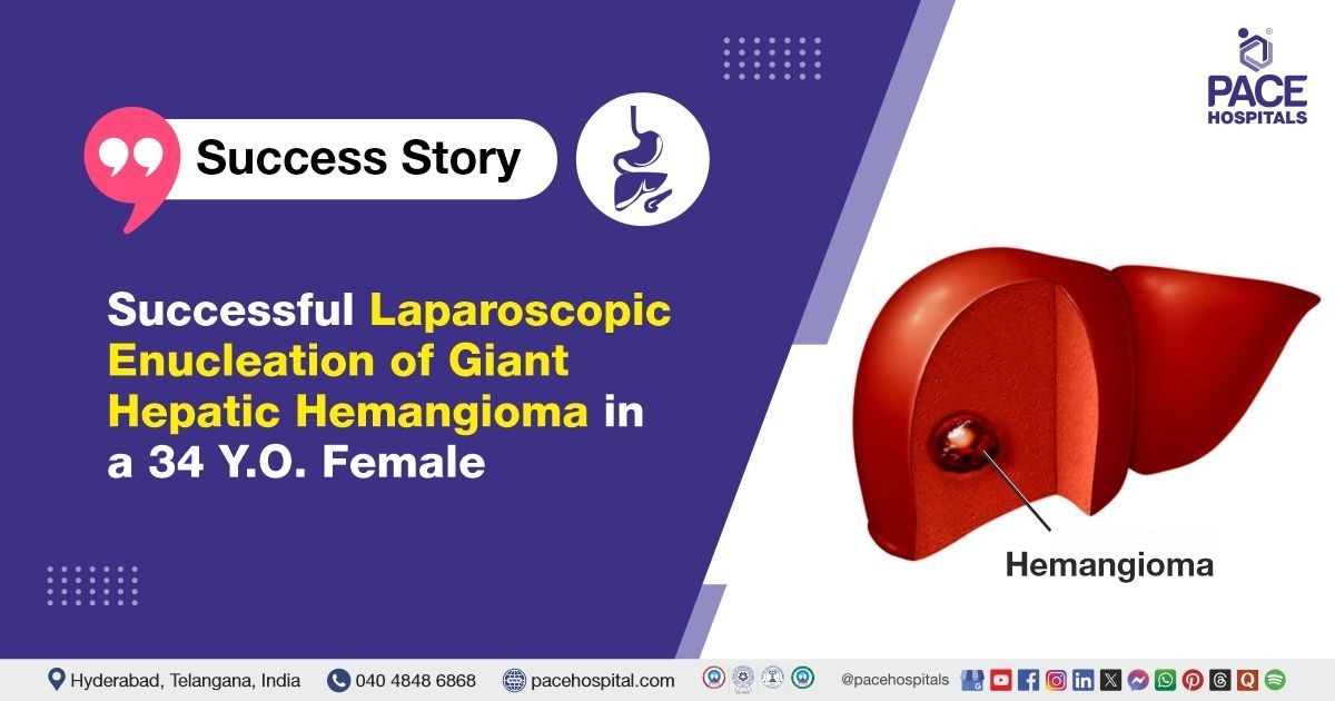
PACE Hospitals’ expert Surgical Gastroenterology team successfully performed a laparoscopic enucleation on a 34-year-old female patient with a hemangioma of the liver involving segments 5 and 8, which was initially suspected to be a space-occupying lesion.
Chief Complaints
A 34-year-old female presented to the Gastroenterology Department at PACE Hospitals, Hitech City, Hyderabad, with complaints of abdominal pain for the past month. She was apparently asymptomatic until a month ago, when she began experiencing a dull, dragging pain in the right hypochondrium (upper right section of the abdomen).
Upon further evaluation, she was found to have a space-occupying lesion in the liver (lump in the liver), with a possibility of haemangioma (a benign blood vessel tumor) involving segments 5 and 8 of the liver. This diagnosis was subsequently confirmed through contrast-enhanced computed tomography (CECT). She then visited the PACE Hospitals for further management.
Diagnosis
Upon arrival at PACE Hospitals, Hitech City, Hyderabad, the patient's vital signs were within normal limits. A thorough physical examination revealed abdominal tenderness localized to the right hypochondrium.
Diagnostic investigations, including contrast-enhanced computed tomography (CECT), revealed a space-occupying lesion (SOL) in the liver, primarily involving segments V and VIII. The lesion was suspected to be a hepatic hemangioma—a common benign vascular tumor of the liver. While often asymptomatic, larger haemangiomas can cause discomfort or a sense of fullness, particularly in the right lobe of the liver. In this case, the lesion’s size and location correlated with the patient’s symptoms.
To confirm the diagnosis, a special scan was done, revealing typical signs of a liver hemangioma, including bright spots around the edges of the lump that gradually filled in toward the center over time.
Based on the diagnosis, she was scheduled for
Laparoscopic Enucleation Surgery in Hyderabad, India at PACE Hospitals, Hitech City, under the expert care of the Surgical Gastroenterology Department.
Medical Decision Making
Following a detailed consultation with Dr. Suresh Kumar S, Consultant Surgical Gastroenterologist, it was determined that a laparoscopic enucleation would be the most appropriate treatment approach for the patient.
Surgical Procedure
Following the confirmed diagnosis, the patient was scheduled for laparoscopic enucleation Surgery at PACE Hospitals, Hitech City, Hyderabad, under the expert care of the Surgical Gastroenterology Department.
After thorough clinical evaluation and preparation, the patient underwent surgery. Intraoperative findings revealed a giant hepatic haemangioma measuring approximately 10×8×6 cm, primarily involving segments V and VIII of the liver. The lesion was exophytic, extending outward from the liver's surface. Given its size and location, the haemangioma posed a risk for potential symptoms and complications, thereby requiring surgical intervention.
The procedure began with Calot’s dissection to clearly identify the biliovascular structures. During the dissection, a feeding artery to the hemangioma was found to arise from the right hepatic artery. This vessel was carefully isolated, clipped, and divided to control the lesion’s blood supply, thereby facilitating safer resection and minimizing intraoperative blood loss.
The liver transection plane was precisely marked, and parenchymal dissection was carried out using a combination of CUSA (Cavitron Ultrasonic Surgical Aspirator) and Ligasure. These tools enabled meticulous and controlled tissue separation. Along the transection line, small hepatic vein tributaries were encountered; each was carefully identified and secured with surgical clips to achieve effective hemostasis and prevent postoperative bleeding.
The specimen, which included both the hepatic hemangioma and the gallbladder, was removed intact. It was extracted through a small Pfannenstiel incision, ensuring minimal tissue manipulation and preserving the integrity of the specimen for histopathological evaluation.
To conclude the procedure, a drain was placed in the subhepatic space to monitor for any postoperative bleeding or bile leakage. This ensured effective drainage, early detection of potential complications, and supported optimal postoperative recovery.
Postoperative Care
The patient’s postoperative recovery was smooth and uneventful, with no complications observed. On postoperative day 3 (POD-3), an ultrasound screening was performed, which revealed normal findings, indicating satisfactory healing and overall recovery.
Discharge notes
The patient made a satisfactory postoperative recovery and was discharged in a stable hemodynamic condition, with the drain still in place. Upon discharge, the following instructions were provided to ensure proper recovery and prevent complications.
Discharge Medications
Upon discharge, the patient was prescribed antibiotics, proton pump inhibitors (PPIs), opioids, and non-steroidal anti-inflammatory drugs (NSAIDs). The patient was also advised to continue with a normal diet as part of the recovery process.
Emergency Care
The patient’s guardians were instructed to seek immediate admission to the Emergency ward of PACE Hospitals, Hyderabad, if they observe any symptoms such as fever, abdominal pain, or vomiting.
Review and follow-up notes
The patient was advised to return for a follow-up appointment with Surgical Gastroenterologists in Hyderabad, India, after three days. This visit will include a comprehensive evaluation, wound assessment, and monitoring of postoperative recovery.
The Role of USG Screening in Optimizing Laparoscopic Enucleation for Liver Hemangiomas
A surgical gastroenterologist/surgical gastroenterology doctor considers ultrasound a crucial tool in the preoperative assessment and surgical planning for laparoscopic enucleation of liver hemangiomas, particularly within the Department of Surgical Gastroenterology.
As a non-invasive, real-time imaging modality, USG provides high-resolution visualization that enables surgical gastroenterologists to accurately assess the size, location, and morphological characteristics of the hemangioma in relation to vital hepatic and vascular structures.
This detailed imaging is essential for evaluating the feasibility of a laparoscopic approach and assists in the precise planning of port placement and dissection planes during surgery. Furthermore, USG is instrumental in identifying potential intrahepatic complications such as vascular involvement, satellite nodules, or multiple hemangiomas, allowing the surgical team to develop a safe, individualized operative strategy for optimal outcomes.
Share on
Request an appointment
Fill in the appointment form or call us instantly to book a confirmed appointment with our super specialist at 04048486868
Appointment request - health articles