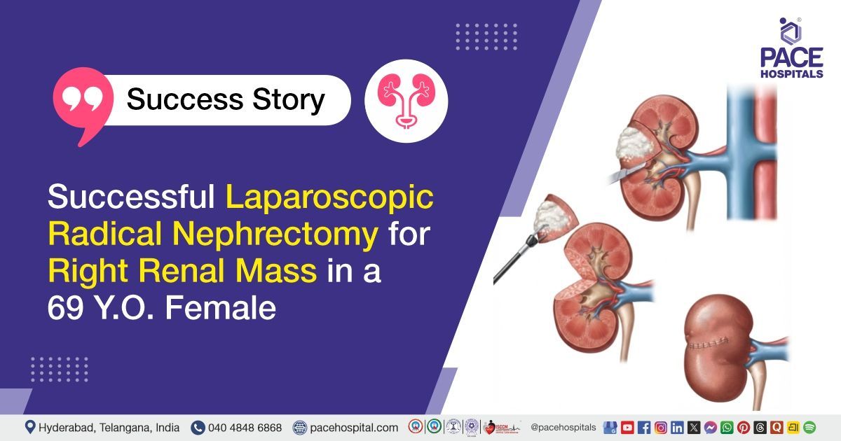
Successful Laparoscopic Radical Nephrectomy for Right Renal Mass in a 69 Y.O. Female
PACE Hospitals
PACE Hospitals’ expert Urology team successfully performed a Right Laparoscopic Radical Nephrectomy on a 69-year-old female patient diagnosed with a right renal mass, along with anemia and diabetes mellitus. The procedure aimed to remove the affected right kidney with the renal mass, support effective disease management, reduce the risk of further complications, and improve the patient’s overall health and quality of life.
Chief Complaints
A 69-year-old female patient with a Body Mass Index (BMI) of 20 presented to the Urology Department at PACE Hospitals, Hitech City, Hyderabad, with complaints of vague right flank pain and generalized weakness. She had no history of hematuria.
Past Medical History
The patient was diagnosed with a right renal mass, clinically staged as cT3. She also had anaemia and diabetes mellitus, which were considered during preoperative evaluation and treatment planning.
On Examination
On examination, the patient was conscious, coherent, and oriented. She appeared weak and had clinical features consistent with anemia. Abdominal and systemic examinations were performed as part of the urology evaluation.
Diagnosis
Upon admission to PACE Hospitals, the patient was thoroughly evaluated by the Urology team, including a detailed review of her symptoms, medical history, and clinical examination. She presented with vague right flank pain and generalized weakness, with no history of hematuria.
The patient underwent relevant investigations, including computed tomography urography, complete blood picture, urine culture and sensitivity, liver function tests, and chest X-ray as part of the preoperative evaluation. Imaging evaluation confirmed the presence of a right renal mass, clinically staged as cT3.
Based on the clinical findings and diagnostic investigations, the patient was diagnosed with a right renal mass, along with anemia and diabetes mellitus. The anemia was considered during preoperative optimization, and diabetes management was included in the overall treatment plan.
Based on the confirmed diagnosis, the patient was advised to undergo Right Renal Mass Treatment in Hyderabad, India, under the expert care of the Urology Department.
Medical Decision-Making (MDM)
After a detailed consultation with Dr. Abhik Debnath (Consultant Laparoscopic Urologist), along with Dr. K. Ravichandra (Consultant Laparoscopic Urologist), a comprehensive evaluation was conducted based on the patient’s complaints of vague right flank pain and generalized weakness. The patient had no history of hematuria, but imaging evaluation revealed a right renal mass, clinically staged as cT3.
The patient underwent required investigations, including computed tomography urography, complete blood picture, urine culture and sensitivity, liver function tests, chest X-ray, and pre-anaesthesia assessment. Her associated anemia and diabetes mellitus were also considered during preoperative planning to optimize surgical safety.
Based on the clinical condition, imaging findings, and diagnosis of right renal mass, it was determined that Right Laparoscopic Radical Nephrectomy was the most appropriate surgical treatment. The procedure helps to remove the affected kidney with the renal mass, reduce the risk of further disease progression, and support better long-term management.
The patient and family members were counselled regarding the diagnosis, need for surgery, laparoscopic approach, possible risks and complications, expected recovery, postoperative care, biopsy review, and the importance of follow-up after surgery.
Surgical Procedure
Following the decision, the patient was scheduled to undergo Right Laparoscopic Radical Nephrectomy Surgery in Hyderabad at PACE Hospitals under the expert care of the Urology Department.
The procedure involved the following steps:
- Patient Preparation and Anaesthesia: The patient was taken to the operating room, and general anaesthesia was administered. Standard aseptic preparation and draping were done, and the patient was positioned appropriately for right-sided laparoscopic kidney surgery.
- Laparoscopic Access and Exploration: Small laparoscopic ports were placed to access the abdominal cavity. The right kidney region was carefully inspected, and the surgical team assessed the renal mass and surrounding structures.
- Mobilisation of the Right Kidney: The tissues around the right kidney were carefully dissected to mobilise the kidney. Nearby structures were protected throughout the procedure, and careful hemostasis was maintained.
- Control of Renal Blood Vessels: The renal artery and renal vein supplying the right kidney were identified, secured, and divided safely to control blood flow to the affected kidney.
- Removal of the Right Kidney with Renal Mass: The right kidney containing the renal mass was removed laparoscopically as part of a radical nephrectomy. The specimen was retrieved safely for further histopathological examination.
- Hemostasis and Closure: The surgical area was checked for bleeding, and complete hemostasis was confirmed. The laparoscopic ports were removed, and the incision sites were closed in layers with a sterile dressing applied.
Postoperative Care
Postoperatively, the patient was closely monitored for pain, bleeding, wound condition, urine output, kidney function, blood sugar levels, and signs of infection. Her recovery was satisfactory, and the Foley catheter was removed on the third postoperative day.
The patient’s haemoglobin improved. Urine culture later showed Klebsiella pneumoniae infection, for which appropriate antibiotics were started. Kidney function and overall health were monitored regularly, and the patient was discharged in hemodynamically stable condition.
Discharge Medications
Upon discharge, the patient was prescribed medicines for infection control, pain relief, fever control, nutritional support, diabetes management, and other supportive care as required.
Advice on Discharge
The patient was advised to continue routine medications, follow a normal diet, maintain home ambulation, and drink 2–3 litres of fluids per day as advised. She was also instructed to do chest physiotherapy, deep breathing exercises, steam inhalation, and incentive spirometry. She was advised to avoid weightlifting and bending forward during the recovery period.
Emergency Care
The patient was instructed to contact the emergency ward at PACE Hospitals in case of the development of symptoms like fever, abdominal pain, vomiting, burning urination, blood in urine, reduced urine output, wound redness or discharge, breathing difficulty, severe weakness, or any sudden deterioration in health.
Review and Follow-Up Notes
The patient was advised to return for follow-up with the Urologist in Hyderabad at PACE Hospitals after 2 weeks for dressing assessment, skin staple removal, and biopsy review.
Conclusion
This case highlights the successful management of a right renal mass with anaemia and diabetes mellitus through right laparoscopic radical nephrectomy. The patient recovered well after surgery, postoperative infection was managed appropriately, and she was discharged in stable condition with medications, recovery advice, and follow-up instructions.
Importance of Timely Surgical Management for Renal Mass
Management of a renal mass depends on its size, location, clinical stage, symptoms, kidney function, and the patient’s overall health condition. In cases where imaging suggests a large or advanced renal mass, timely evaluation by a urologist/urology doctor is important to determine the most suitable treatment approach.
Laparoscopic radical nephrectomy is commonly considered when the affected kidney needs to be removed completely to manage the renal mass effectively. Proper preoperative assessment, including CT imaging, blood tests, urine tests, kidney function evaluation, and anaesthesia fitness, helps improve surgical safety. Associated conditions such as anemia and diabetes also require careful monitoring before and after surgery.
Postoperative care focuses on pain control, wound healing, infection management, kidney function monitoring, breathing exercises, early mobilisation, and follow-up biopsy review. A planned and patient-centred approach helps support safe recovery and long-term health after kidney surgery.
Frequently Asked Questions (FAQs)
What is a renal mass?
A renal mass is an abnormal growth in or on the kidney. Some renal masses may be benign, while others may be cancerous, so proper evaluation is important. Imaging tests such as ultrasound, CT scan, or MRI help doctors understand the size, location, and nature of the mass before planning treatment.
Why can a renal mass cause flank pain?
A renal mass can cause flank pain when it increases in size, stretches the kidney covering, presses on nearby tissues, or affects urine drainage. Some patients may have only vague pain or weakness, while others may not have any symptoms at all. This is why imaging tests are important when persistent flank pain is present.
Why is the entire kidney removed in radical nephrectomy?
The entire kidney may be removed when the mass is large, advanced, or not suitable for kidney-sparing surgery. In radical nephrectomy, the affected kidney is removed to manage the disease effectively and reduce the risk of spread or recurrence. The decision depends on the tumour size, location, stage, and the patient’s overall health.
How is anemia related to kidney disease or kidney tumors?
The kidneys help produce erythropoietin, a hormone that supports red blood cell production. When kidney function is affected, red blood cell production may reduce, leading to anaemia. In some patients with kidney tumours, anaemia may also occur due to chronic illness, bleeding, inflammation, or poor nutritional status.
How is urinary infection treated after kidney surgery?
A urinary infection after kidney surgery is usually treated with antibiotics selected according to urine culture and sensitivity results. Doctors may also monitor fever, urine output, pain, blood tests, and kidney function. Completing the prescribed treatment and attending follow-up is important to prevent the infection from worsening.
Why are breathing exercises advised after laparoscopic surgery?
Breathing exercises, incentive spirometry, and chest physiotherapy help expand the lungs after surgery. They reduce the risk of chest congestion, mucus retention, and postoperative lung infection, especially after general anaesthesia. These exercises also support better oxygen levels and smoother recovery.
What tests help identify whether a renal mass is serious?
Imaging tests such as CT urography, CT scan, MRI, or ultrasound help identify the size, location, spread, and features of a renal mass. Blood tests, urine tests, chest imaging, and kidney function tests may also be done before surgery. In some cases, biopsy or histopathology after removal confirms whether the mass is benign or cancerous.
What is the difference between partial nephrectomy and radical nephrectomy?
Partial nephrectomy removes only the tumor or diseased part of the kidney while preserving as much healthy kidney tissue as possible. Radical nephrectomy removes the entire affected kidney and sometimes nearby tissues, depending on the extent of the disease. Partial nephrectomy is usually preferred when safely possible, while radical nephrectomy may be needed for larger or more advanced masses.
Is laparoscopic nephrectomy better than open nephrectomy?
Laparoscopic nephrectomy is a minimally invasive approach done through smaller incisions, and it may lead to less pain, shorter hospital stay, and faster recovery in suitable patients. Open surgery may still be preferred for very large, complex, or difficult tumours. The best approach depends on the patient’s condition, tumour features, and surgeon’s assessment.
What diet is advised after kidney removal surgery?
After kidney removal surgery, patients are usually advised to follow a balanced diet, drink adequate water as recommended, and avoid excess salt. Protein intake should be appropriate and guided by kidney function and doctor’s advice. Patients with diabetes should also follow blood sugar-friendly food choices to support wound healing and protect the remaining kidney.
Share on
Request an appointment
Fill in the appointment form or call us instantly to book a confirmed appointment with our super specialist at 04048486868
Appointment request - health articles
Recent Articles