Successful Paediatric Liver Transplantation (PLT) for a 7-Month-Old Baby Girl with Biliary Atresia | Case Study
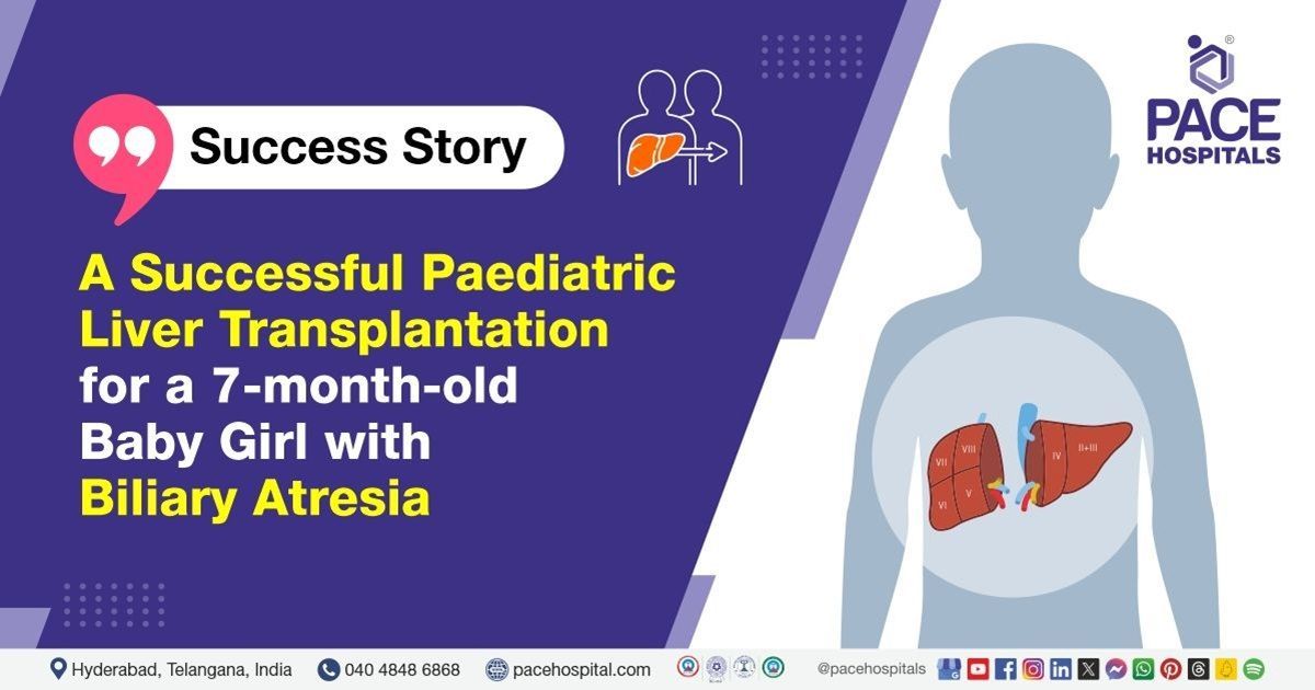
Liver Transplantation team at PACE Hospitals successfully performed Paediatric liver transplantation (PLT) in a 7-month-old infant suffering from congenital biliary atresia with secondary biliary cirrhosis and managed postoperative complication of jejunal perforation.
A baby girl of 7 months old presented to the PACE Hospitals with complaints of
jaundice for the last six months, which showed a progressive and gradual development of bilirubin levels.
Medical History
Delving deeper, it was understood that the baby girl had been experiencing jaundice for the last six months. She was a known case of extrahepatic biliary atresia type 1 (partial or total absence of the permeable bile duct between the porta hepatis and the duodenum) as evidenced by the presence of jaundice, clay-colored stool, and hepatomegaly (increased liver size) early in her life.
Consequently, around four months ago, she underwent Kasai’s procedure (also called Kasai hepatoportoenterostomy), a surgery to treat biliary atresia. At the time of Kasai’s procedure, it was reported that the patient had the following:
- Gross hepatomegaly accompanied by hard consistency of the liver tissue
- Neovascularisation (formation of new blood vessels) was seen
- Chronic cholecystitis was seen (swelling and irritation of the gallbladder)
Diagnosis
Upon being admitted to PACE Hospitals and reviewing the medical history, the patient was subjected to
investigations including
liver biopsy and blood tests. Evaluating the diagnostic results, the patient was diagnosed with Congenital Biliary Atresia Type-I with Secondary Biliary Cirrhosis. The Paediatric End-Stage Liver Disease (PELD) score is 30.
Treatment
Liver transplantation team of Dr. CH Madhusudhan, Dr. Ravula Phani Krishna, Dr. Govind Verma and Dr. Suresh Kumar asserted that a liver transplant was the only way to salvage / save the patient.
The patient was kept on multiple lifesaving supports. With necessary investigations done & clearances obtained, which included a pre-anesthesia checkup, the patient underwent Paediatric liver transplantation (PLT), receiving the left lateral segment. The procedure was supervised by the liver transplant surgeon Dr. CH Madhusudhan, and it was accomplished devoid of any complications.
The aftermath
The post-operative period was uneventful which can be evidenced by the results of liver doppler test (a non-invasive test which studies the blood supply in and around the liver through ultrasound waves).
Five days after the transplant the ultrasonography screening was done which showed a collection of fluid in left lumbar and right sub diaphragmatic space. To drain the fluid, Percutaneous transhepatic biliary drainage (PTBD) insertion was done, thus relieving the pressure.
On the sixth day post-surgery, the subject vitals monitoring devices showed tachycardia (increased heart rate), abdominal distension, and gaurding. Since all these symptoms indicate the general direction of suspecting hollow viscus perforation, the surgeons planned for an emergency exploratory laparotomy. Indeed, they found jejunal perforation – a very rare case, especially after liver transplantation. The jejunum is the middle part of the small intestine. Through emergency exploratory laparotomy, they could close the primary jejunal perforation.
The necessary medicines, immunosuppressives, antibiotics, proton pump inhibitors, multivitamins, antiemetics, analgesics, antipyretics & other supportive care were given along with the counseling. Once the patient achieved hemodynamic stabilisation, she was discharged with the necessary medications and advice for follow-up.
Jejunal perforation – A mortal complication of liver transplantation
Jejunal perforation is an extremely rare but potentially mortal complication that occurs in the jejunum of the gastrointestinal tract.
While a delayed diagnosis can certainly be life-threatening, it must be understood that an early diagnosis is challenging due to the expression of a similar set of atypical clinical features, which are commonly seen with large doses of steroids and immunosuppressants. Necessarily, large doses of steroids and immunosuppressants are often given in liver transplant cases, and it takes medical and surgical expertise to correctly identify and diagnose the features.
The cause of jejunal perforation after liver transplant cases could vary and the possible risk factors include previous laparotomy, prolonged surgery, subsequent laparotomy, portal vein thromboembolism, treatment with high-dose steroids, and cytomegalovirus (CMV) infection.
The incidence of jejunal perforation after liver transplant is 1-5.3% in adults and 8.3-14% in children. The higher incidence in children is most likely attributable to tight adhesions of the liver and the formation of intestinal loops during portoenterostomy before liver transplant. Nevertheless, a Kasai’s procedure (portoenterostomy procedure) which was performed before liver transplant in children appears to be a serious risk factor.
Share on
Request an appointment
Fill in the appointment form or call us instantly to book a confirmed appointment with our super specialist at 04048486868

