
Thoracic Disc Herniation - Symptoms, Causes, Preventions, Treatment
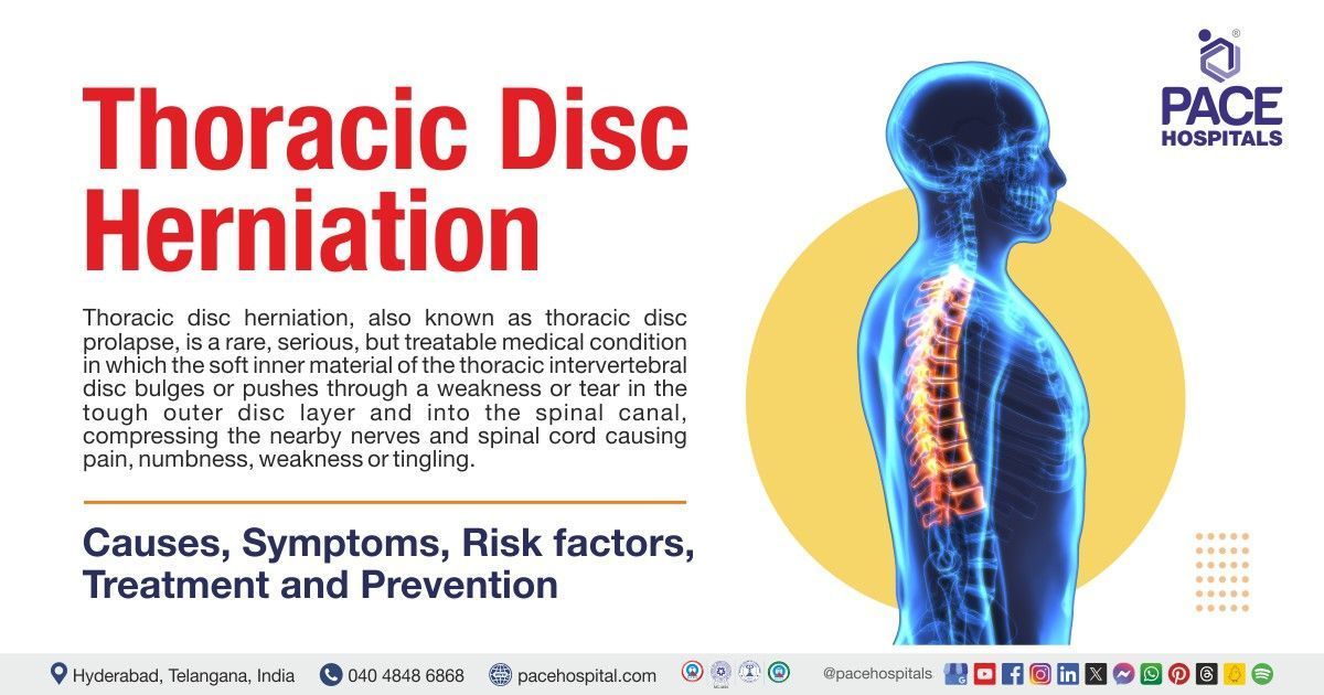
Thoracic disc herniation, also known as thoracic disc prolapse, is a rare, serious, but treatable medical condition in which the soft inner material of the thoracic intervertebral disc bulges or pushes through a weakness or tear in the tough outer disc layer and into the spinal canal.
The protruding or bulging disc material may put pressure on the nearby nerves of the thoracic region and spinal cord, causing upper back and chest pain, discomfort, numbness, or tingling.
The thoracic spine has 12 vertebrae (bones), numbered T1 to T12, starting from the base of the neck (upper back) down to the abdomen (mid-back); these bones are interlocked in the spinal column and attached to a rib on either side, and the thoracic vertebrae is the only spinal section which is attached to the rib cage, where it provides support to the thoracic spine, making it stronger and less likely to experience injury (wear and tear) than the other sections of the spine.
Prevalence of thoracic disc herniation
It is uncommon, with an occurrence percentage of 0.5 to 4.5 of all disc ruptures. It's usually discovered accidentally through MRI scans. Also, it rarely causes noticeable symptoms, accounting for only 0.25% to 0.75% of cases. Approximately 63% of those affected show symptoms, and the overall occurrence of this condition is quite rare, about one in a ten lakh cases.
Surgeries for thoracic disc herniation cases are relatively rare compared to other parts of the spine, such as cervical and lumbar, accounting for only about 0.15% to 1.8% of herniation cases and most commonly affecting individuals between 40 and 60 years old.
Women and men might be affected in equal proportions. Most individuals, about 80%, usually experience herniation issues in 30s and 40s. Around 75% have this issue below the eighth thoracic vertebrae (T8), with the highest incidence between the T11 and T12 regions.

Thoracic disc herniation causes
In some cases, the cause of thoracic disc herniation is unknown (idiopathic). However, the possible thoracic disc herniation causes include the following:
- Age: Thoracic disc herniation most often occurs due to the gradual wear and tear that comes with aging. As the person ages, the cushion-like discs between the vertebrae (bones) weaken, resulting in the inner part of the disc bulging out through the outer layer. This may put pressure on nearby nerves or the spinal cord, which may cause symptoms such as shooting pain along nerves (radicular) or issues with sensation and movement (myelopathic).
- Degenerative disc disease: It is the process in which a cushion-like disc between the vertebrae loses hydration and becomes weak. The exact cause of disc degeneration is considered multifactorial; several factors such as injuries, metabolic irregularities, genetics, vascular (blood flow problems), and infections may contribute to the wear and tear of the disc. Among these factors, injuries or trauma happen to be one of the most common causes of this condition.
- Trauma or injury to the upper back: Traumas or injuries such as accidents or sudden falls may force the thoracic spine to cause a tear as a result the disc's inner gel bulges through the tougher outer layers.
- Strain: Strain, associated with repeated axial spine twisting (rotational movement along the spinal axis),causes spinal instability and changes in alignment. These changes may increase the risk of developing disc degeneration (wear and tear), raising the chances of thoracic disc degeneration in the long run.

Thoracic herniated discs symptoms
Thoracic disc prolapse symptoms may range from mild to severe, where the thoracic spinal cord is narrower than the neck and lower back, and even slight compression may cause severe symptoms. However, it rarely compresses nearby nerves because of limited mobility and may cause localized pain, discomfort, burning sensations, or even difficulty breathing if it affects specific nerves.
Thoracic spine herniated disc symptoms often include the following:
- Radiculopathy
- Myelopathy
Thoracic disc herniations may cause different types of pain, including radicular and myelopathic pain, based on whether the herniated disc compresses the nerve roots or the spinal cord.
Radiculopathy: The initial common symptom of a herniation is mid-back pain (upper back pain), usually in the center, which may radiate to one or both sides depending on the location and severity of the herniation. Radicular pain due to nerve root compression that follows specific paths on the body, such as
- T1 pain that radiates to the inner forearm
- T2 pain that radiates to the armpit
- T4 pain that radiates to the nipple areas
- T10 pain that radiates to the belly button
- T12 pain that radiates to above the groin
These symptoms of a herniated disc in the upper thoracic spine may allow neurosurgeon or orthopaedic surgeon to find which disc is causing the herniation based on where the pain is felt. However, the pain is not continuous, and it might comes and goes, worsens if the patient strains or coughs. Sometimes, this pain might even spread to the sides or legs.
Myelopathy: Myelopathic pain caused by spinal cord compression and shows various symptoms such as:
- Numbness or tingling
- Weakness and pain in the lower body
- Hyperreflexia (overresponsive or overactive bodily reflexes to stimuli), Increased reflexes in legs
- and spasticity (muscle stiffness)
- Gait abnormalities: Unusual walking pattern (Issues in walking)
- Bowel and bladder dysfunction
- Rarely paraplegia: Paralysis that occurs in the lower half of the body
Many people with thoracic disc herniation might not show noticeable symptoms (asymptomatic) or present with non-specific symptoms, including chest wall pain, upper extremity pain or epigastric pain, and rarely, pain in the groin or the lower extremity, which creates a suspicion of other common conditions, because of diverse symptomatology and infrequent occurrence of thoracic disc herniation.

Thoracic disc herniation risk factors
Various factors may contribute to developing a herniated thoracic disc. Common risk factors for developing thoracic disc herniation include as follows:
- Age: Weakening of the disc happens with age, so middle-aged individuals -are more prone to disc problems.
- Diseases of the thoracic spine: Thoracic spine diseases, including degenerative disc trauma or disease leading to weakening of discs. This weakening may increase the risk of developing thoracic disc herniation.
- Being overweight: Being overweight may put excess pressure on the person's spine, leading to wear and tear on the thoracic discs, resulting the increased risk for developing thoracic disc herniation.
- Poor Posture: Poor posture may increase strain, pressure and reduces flexibility on the spine, which may affect the thoracic disc, making it more vulnerable to herniation.
- Joint stiffness: Muscle stiffness may put more strain during movement due to limited flexibility and mobility, which may increase the chances of disc herniation in the thoracic region.
- A sedentary lifestyle: Lack of exercise may weaken the muscles that support the spine, potentially raising the risk of cervical disc herniation.
- Scheuermann's disease (abnormal rounding of the upper back vertebrae in adults):
Individuals with Scheuermann's disease (a spine condition) are more prone to get a thoracic herniated disc.

Thoracic disc herniation complications
Thoracic disc herniation complications may arise from an untreated thoracic herniated disc. The thoracic area has less movable larger discs, but due to the presence of ribs they are less prone for herniation. However, they are more susceptible to developing complications if they occur. The following are the complications of thoracic disc herniation:
- Complete paralysis below the waist
- Damage to the nerves in the lower body and legs
- Spinal cord injury
Most cases of thoracic disc herniations go through calcification, making them harder, increasing the strain and pressure on the spinal cord and worsens symptoms. Sometimes, the calcified disc may stick or tear the dura (the protective layer of the spinal cord), leading to a leak of cerebrospinal fluid.
- Cerebrospinal fluid leak: If this happens, patients might experience unusual symptoms such as:
- Intracranial pressure issues: Pressure caused by cerebrospinal fluids inside the brain tissue and skull, which is a dangerous and emergency condition
- Postural or orthostatic hypotension: Sudden drop of blood pressure (low blood pressure) when a person stands up
- Headaches
Thoracic herniated disc diagnosis
Diagnosis of thoracic disc herniation mainly includes the following:
- Medical history
- Physical examination
- Imaging tests
- X-rays
- Magnetic Resonance Imaging (MRI)
- Computed Tomography (CT) imaging
- CT myelography
- Other tests
- Electrical tests (EMG and SSP)
Unlike common cervical and lumbar disc herniations, most thoracic disc problems are often asymptomatic (don't show symptoms) and found incidentally on MRI. When symptoms do appear, they are frequently unusual and not associated with the spine, making diagnosis difficult, and there is a chance of ruling out other common conditions.
The initial step in diagnosing a thoracic herniated disc includes a patient's medical history and physical examination to find the symptoms, such as pain and its location.
Medical history
The neurosurgeon or orthopaedic surgeon (or primary care physicians, pain specialists, chiropractors, physical therapists) first asks the questions regarding the patient's medical history to identify any pre-existing conditions, accidents, or trauma that may have caused an injury to the individual spine prior to the thoracic back pain or any other conditions, such as fevers, weight loss, difficulty urinating.
Diagnosing thoracic disc herniations is tricky because symptoms may resemble other health issues such as lung, heart, stomach, or urinary problems rather than typical neck or back pain. Unlike lumbar or cervical problems that frequently cause leg or arm pain, thoracic disc herniations may have vague symptoms, making them hard to detect. In some cases, lateral herniation around C7-T1 or T1-2 can cause Horner's syndrome, with specific eye and facial symptoms.
Hence, it is always essential to find out the other conditions that may be causing the radicular pain and mimic it, mainly including:
- Diabetes
- Shingles
Mechanical causes:
- Muscle strain
- Rib or facet joint fractures
- Issues with collarbone
These two conditions present with distinct signs like skin rashes for shingles and abnormal metabolism for diabetes.
Physical examination
A neurosurgeon or orthopaedic surgeon (including nurse practitioners, primary care physicians, pain specialists, chiropractors, physical therapists) may start a physical exam to understand better the patient's symptoms by touching and pinpricking certain areas to check sensation and evaluate for radiculopathy in the upper body and myelopathy in the lower body.
Initially, a neurosurgeon or orthopaedic surgeon assesses the following:
- Location of the pain
- The severity of the pain
- Type of pain (burning, weakness, numbness, etc.)
Additionally, for the lower body, a neurosurgeon or orthopaedic surgeon may assess
- Reflexes (to check abnormal reflex responses),
- Proprioception (ability to sense movement and position of limbs without looking) and
- Muscle tone (amount of tension or resistance in muscles) might be evaluated to gauge nerve function and spinal cord health.
Imaging tests:
Imaging tests might be recommended to diagnose the herniation accurately and evaluate the extent of the condition.
- X-ray: A neurosurgeon or orthopaedic surgeon (including primary care physicians, radiologists, pain specialists, or physical therapists) may recommend taking X-rays of the middle back. Regular X-rays may not show a herniated disc, but they may show (give an idea) the amount of wear and tear happened in the spine and may show other causes. However, X-rays are not as precise as MRI for diagnosing a herniated thoracic disc.
- Magnetic resonance imaging (MRI): Thoracic disc herniations (TDHs) are uncommon and might not exhibit typical symptoms, leading to delayed diagnosis. However, MRI scans are significantly helpful for diagnosing TDHs, as they provide highly detailed pictures that assist neurosurgeons or orthopaedic surgeons (including primary care physicians, orthopaedic and neurosurgeons, radiologists, pain specialists, physical therapists) in precisely spotting and confirming these rare spine problems. It is the common test for thoracic herniated disc diagnosis it and is the best technique for diagnosing and observing issues with intervertebral discs. This test is very accurate and painless, with no side effects. However, sometimes, the MRI cannot interpret the complete cause. Therefore, other tests might be recommended.
- Computed Tomography (CT) imaging: It is a highly sensitive test for evaluating the bone structures of the spine, revealing details about degenerative changes or fractures. This test can also identify bone-related issues or calcified herniated discs that do not readily appear or are visible on other scans. In patients unable to undergo MRI testing, CT myelography is an alternative method to display herniated discs.
- CT myelogram: A myelogram, usually combined with a CT scan, may be required to give as much information as possible.
CT myelogram is a vital imaging method that combines the benefits of myelography and the high resolution of CT. It utilizes a contrast dye and X-rays or computed tomography (CT) to check problems in the spinal canal, spinal cord, nerve roots, and other tissues. The radiologist may remove some amount of spinal fluid from the spinal canal and inject a small amount of contrast dye, and the X-ray table might be tilted in different directions to pass the contrast dye to various areas of the spinal cord to get detailed images of the body.
Other tests:
A neurosurgeon or orthopaedic surgeon may suggest other tests when the imaging tests do not provide detailed information about the causes of a thoracic herniated disc.
- Electrical tests (EMG and SSP):
These tests are performed to confirm the pain in the leg is coming from a damaged nerve.
Thoracic herniated disc treatment
Herniated disc treatment in thoracic spine is categorized into three types, namely:
- Conservative treatments
- Interventional treatments
- Surgical treatment
Conservative treatment:
- Observation
- Rest
- Medications
- Pain medications: Narcotic and non-narcotic analgesic medications
- Chiropractic manipulation
- Physical activity
Interventional treatments:
- Anti-inflammatory injections: Epidural steroid injection (ESI)
Surgical treatment:
- Laminotomy and discectomy
- Microdiscectomy
- Costotransversectomy
- Transthoracic decompression
- Thoracic fusion
- Video-assisted thoracoscopy surgery (VATS)
Conservative treatment
One of the treatment options for this condition is conservative management, which includes over-the-counter analgesics, non-steroidal anti-inflammatory medication, and physiotherapy, which involves strengthening through extension exercises.
Thoracic spine herniated disc treatment depends on the symptoms, such as whether the symptoms are getting better or worse. If the symptoms get worse, surgery might be recommended, or if the symptoms are improving, watching and waiting (observation) might be suggested to see whether the symptoms reduced, and conservative treatment of thoracic disc herniation includes the following:
- Observation: Many cases may completely resolve within several weeks or months. A patient may not need treatment other than watching to ensure the issue does not progress. If the patient is bearing the pain and there is no progression of weakness or numbness, a neurosurgeon or orthopaedic surgeon may suggest to watch and wait.
- Rest: If the herniated disc pain is more severe, a patient may be advised to take a few days off from work and reduce the activity for a while. After several days, the patient might be suggested to begin gentle walking and increase the walking distance each day.
- Pain medications: Based on the severity of pain, multiple approaches may be used to help control the patient's pain with medications. It includes OTC (over the counter) and some anti-inflammatory drugs. If these medications are not able to control the pain, a neurosurgeon or orthopaedic surgeon may prescribe some stronger pills for pain, such as narcotic or non-narcotic pain medications.
- Narcotic pain medications may be prescribed to reduce the pain, but they are addictive and very strong
- Non-narcotic pain medications are somewhat less effective than narcotics
However, most neurosurgeons or orthopaedic surgeons prescribe narcotics for more than a few days or weeks based on the patient's condition.
- Chiropractic manipulation: Chiropractic manipulation is a treatment where a trained neurosurgeon or orthopaedic surgeon practitioner called a chiropractor either uses a small instrument or hand to apply a controlled, sudden pressure and force to a spinal joint to restore and improve the motion of the spine and physical function and to ease the pain.
- Physical activity: A physical therapist may direct a rehabilitation program for patients referred by a neurosurgeon or orthopaedic surgeon. This program aims to control symptoms, find positions that relieve pain, improve back movement and posture, and teach the patient how to keep the spine safe during everyday activities.
Physical therapy for thoracic herniated discs may design a rehabilitation program based on the patient's condition to prevent future problems. As patients recover, they gradually perform a sequence of strengthening exercises, such as walking or swimming (aerobic exercises), to alleviate pain and improve endurance. However, the patient has to gradually advance the thoracic disc herniation exercises that the therapist might recommend to avoid the surgery. Thoracic herniated disc exercises to avoid includes jumping, running, jogging, squats, and leg presses, where these activities may cause repetitive loading and strain on the patient's lower back.
Interventional treatments:
A neurosurgeon or orthopaedic surgeon might recommend interventional treatments for a thoracic herniated disc to mainly reduce the pain, inflammation, and other symptoms that do not respond to non-surgical approaches.
- Epidural steroid injection (ESI): Usually, ESIs are used for severe pain from a herniated disc, when surgery is near. They may not be the first choice and could have only around 50% success rate. ESIs are usually suggested after exploring other options, with the aim of reducing pain before considering surgery.
Spinal injections are injected under X-ray guidance, also known as fluoroscopy. This allows the neurosurgeon or orthopaedic surgeon to visualize the spine to ensure accurate needle placement.
Surgical treatment
Surgery is mainly indicated when the patient has severe back pain, neurological deficits, or stubborn intercostal neuralgia. Surgical approaches for the herniated thoracic disc include the posterior, posterolateral, ventral, and thoracoscopic approaches.
Calcified hernias at the spine's mid-line might be approached using a transthoracic incision (cutting through the chest wall to see structures in the thoracic area), while lateral soft hernias might be approached using a posterolateral incision (cutting on the back side, to access the lateral or outer regions of the spine). The complication rates are higher for the transthoracic approach; however this approach can be used for complex cases than the posterolateral approach. Fusion surgery is suggested for multiple herniations and Scheuermann's disease-related herniations.
The type of surgical approach depends on the disc size, spine level, disc location, presence of calcification, and overall health condition of the patient. Usually, surgery is indicated for individuals with clear neurological problems and progressive myelopathy.
Other reasons for surgery include ongoing radicular pain and stable myelopathy without major functional problems. Emergency surgery might be required for patients where neurological issues may lead to worsening myelopathy or functional impairment. The following are some of the common surgical approaches that can be used to treat the thoracic disc herniation:
- Transthoracic Decompression: Thoracic decompression is the removal of a complete or partial portion of the bone that covers the back of the spinal column called the lamina, along with removing a part of the rib on the affected area. This surgery aims to access the bone or disc, causing pressure around the nearby nerve or spinal cord.
- Laminotomy and Discectomy: These procedures are the traditional way of treating the thoracic herniated disc. Laminotomy is a medical procedure in which a tiny opening (making an opening in the lamina) is made between the two vertebrae in the region of disc rupture. This allows the neurosurgeon or orthopaedic surgeon to see into the patient's spinal canal and make space to work. Discectomy refers to a medical procedure called "removal of the disc." This is performed to relieve pressure and reduce the irritation on the spine nerves. After laminotomy, the neurosurgeon or orthopaedic surgeon moves the nerve roots away, locates the ruptured disc material, and removes it from the spinal canal.
- Costotransversectomy: It allows for exposure of the costovertebral joints, neural foramina, and lateral spinal cord. In this procedure, the neurosurgeon or orthopaedic surgeon makes a posterolateral incision through the back of the spine to remove the ends of one or more nearby ribs, where they join the spine. A portion of the transverse process (the small bone on the side of the vertebra) is taken off. Hence, this allows the neurosurgeon or orthopaedic surgeon to see the injured disc by forming. Finally, the surgeon may locate and remove the disc ruptured disc material by decompressing the spinal cord.
- Microdiscectomy: Microdiscectomy is similar to the traditional laminotomy and discectomy. However, to perform this, a much smaller incision is needed. This procedure causes less damage to the spine (during the disc surgery) and faster recovery for the patient. The neurosurgeon or orthopaedic surgeon will use the microscope to see through the smaller incision. A tiny incision is made in the back just above the site where the disc is herniated. The rest of the surgery is executed precisely like the traditional laminotomy and discectomy.
- Thoracic Fusion: The surgery is performed to join two or more bones into one solid bone because, in some cases, a degenerated thoracic disc may require to be removed completely and the adjacent bones fused together to stabilize and support the spine. Initially, a neurosurgeon or orthopaedic surgeon removes the damaged or injured thoracic disc, which will be replaced with a bone graft. A bone graft may be taken from the autograft (patient's rib or hip) or the allograft (from a cadaver donor). After the placement of the bone graft, the surgeon may place rods and screws in the bone (vertebrae) above and below the removed disc. This hardware material (Rods, plates, and screws) will help hold the bones in place to heal the bone graft. The bone graft material might be used to help unstable bones to grow together.
- Video Assisted Thoracoscopy Surgery (VATS):
A neurosurgeon or orthosurgeon may remove damaged portions of the disc through a small incision into the side of the thorax, using the thoracoscope while watching the process on a small television camera. This procedure is easier to perform, avoids the formation of scar tissue around the nerves, and helps the patients to recover fast.
Rehabilitation
Rehabilitation programs, including joint stability training and mobilization, might be initiated after surgery. Other programs such as range of motion, low–impact aerobic activity, and extension-focused strengthening exercises are recommended to restore movement (mobility), strengthen the muscles, and improve function.
Some home exercises for abdominal and paraspinal muscles (muscles surrounding and attaching to the spine) and cardiovascular conditioning may be suggested to prevent recurrences.

Thoracic disc herniation prevention
The following are some preventive measures that one may take to avoid thoracic disc herniation:
- Managing weight: Excess weight is the major risk factor for many health conditions; a healthy weight reduces the strain on discs. Therefore, managing weight is essential to prevent spine-related problems such as disc herniations.
- Avoid lifting heavy objects: Lifting the heaving objects suddenly may cause immense pressure on the spine, increasing the risk of disc herniations or displacement. Therefore, neurosurgeons or orthopaedic surgeons suggest avoiding lifting heavy objects suddenly, especially for older people.
- Maintaining a good posture: Poor posture may cause prolonged stress and misalignment to the spine, leading to herniation in the thoracic region. Proper posture techniques help reduce the pressure and strain and prevent the chances of getting herniation problems in the future.
- Exercising: Adding moderate physical activity, such as walking, is advisable as an everyday routine because exercising may strengthen the muscles and improve flexibility.
- Having a proper diet: A nutritious diet is essential to maintain a healthy weight, which is helpful to avoid being overweight. Eating green and leafy veggies, nuts, oily fish, and other foods may help in fight inflammation if present.
- Avoiding prolonged sitting: Avoiding long-time sitting or standing to avoid increased pressure strain on the spinal discs. Doing simple neck stretches while sitting or standing in the same position for a long time strengthens the muscles.
Difference between thoracic and cervical disc herniation
Thoracic vs Cervical disc herniation
Disc herniations in the thoracic and cervical spine are common conditions that share some similarities but also have differences, which include:
| Elements | Thoracic disc herniation | Cervical disc herniation |
|---|---|---|
| Location | Herniated thoracic disc occurs in the upper back and mid region of the spine. | Herniated cervical disc occurs in the neck (cervical) region of the spine. |
| Affected nerves | T1 – T12 | Between C5-C6 and C6-C7 |
| Symptoms | Symptoms mainly include upper back pain, which radiates to chest or abdomen. | Symptoms mainly include neck pain, which extends to shoulder, arm and fingers. Along with pain, some patients may experience numbness or weakness in the upper extremities. |
| Causes | Common causes of thoracic disc herniation include age related degeneration, repetitive stress and injury to the thoracic region. | Usually, it occurs due to degeneration, trauma or injury in the neck region, repetitive neck movements, sudden twisting or bending of the neck |
| Prevalence | Less common than cervical and thoracic disc herniation. | More common than thoracic disc herniation. |
| Complications | Complications mainly include complete paralysis below the waist, damage to the nerves in the lower body and legs, or spinal cord injury. | Complications of herniated cervical disc includes radiculopathy, autonomic dysfunction and hyperreflexia. |
| Treatment | Treatment options include rest, physical therapy, pain medications. | Treatment options include observation, pain medications, physical therapy, neck mobilization and surgery. |
| Surgery | Surgical approaches of thoracic disc herniation mainly include laminotomy and discectomy, microdiscectomy, costotransversectomy, transthoracic decompression, thoracic fusion and video-assisted thoracoscopy surgery (VATS). | Surgical approaches of cervical disc herniation include anterior cervical discectomy and fusion (ACDF), posterior cervical laminoforaminotomy, total disc replacement, cervical corpectomy and fusion. |
Frequently Asked Questions (FAQs) on Thoracic disc herniation
How serious is a thoracic herniated disc?
Herniation in the thoracic region is rare due to less mobility in the vertebrae, which do not provide extra space. However, they are more susceptible to developing complications if herniation occurs. Therefore, when a herniated disc occurs in the thoracic region, a neurosurgeon or orthopaedic surgeon often considers it (herniation) serious because, in severe cases, the pressure on the spinal cord may lead to paralysis below the waist. Fortunately, thoracic herniated discs are not as common as the cervical and lumbar spine.
Can herniated disc lead to paralysis?
Herniated thoracic disc may cause paralysis. However, the onset of paralysis is not sudden and complete; it gradually develops in this condition, along with other symptoms called pain in the thoracic area. Many factors can lead to this condition, mainly acute cervical disc ruptures that cause severe cord compression and make a patient paralyzed. However, many cases of herniated thoracic discs will be diagnosed and treated before they progress to partial paralysis.
How is a thoracic herniated disc treated?
Most thoracic disc herniations may be treated with a conservative treatment (non-surgical approach) that usually includes rest, observation, anti-inflammatory drugs (medications), physical therapy, and the use of supports or back braces (back brace for thoracic herniated disc) to stabilize and support the spine. However, a surgical approach might be recommended if the patient is experiencing worsening symptoms, such as severe back pain and neurological symptoms that are not responding to non-surgical treatment
How to sleep with a thoracic herniated disc?
One can sleep on the either side, curled in a foetal position, to decrease or stop the pain caused by herniated discs. One can attain that position by lying on the back and rolling over the side. Other positions include:
- Side-lying position (lying on the side with a pillow between legs)
- Back lying position (using a pillow under the knees by turning back)
- Reclining position (using a pillow under the head and knees)
These positions help keep the spline aligned and reduce pressure, allowing the person to sleep well.
How common are thoracic disc bulges?
It’s quite common to experience lower back or neck (lumbar or cervical) disc herniations, but a herniation in the thoracic spine region is extremely rare. Herniation in the thoracic region accounts for less than one percent of all herniated discs, which occurs in one out of every one crore people annually. Hence, the chances of herniation are very low, quite literally one in ten lakhs.
The reason for less prevalence is that the thoracic region has more vertebrae than the lumbar and cervical, and they have less mobility and, therefore, are less prone to herniation.
Share on
Request an appointment
Fill in the appointment form or call us instantly to book a confirmed appointment with our super specialist at 04048486868
Appointment request - health articles


Our Locations – Find the Best Hospital Near You
Metro Pillar Number C1772, Beside Avasa Hotel, Hitech City Road, Near HITEC City Metro Station, Hyderabad, Telangana, India.
Mythri Nagar, Beside South India Shopping Mall, Hafeezpet, Madeenaguda, Hyderabad, Telangana, India.
040 4848 6868
Payment in advance for treatment at PACE Hospitals, Hyderabad, Telangana, India (Pay in INR ₹)
For Bank Transfer:-
- Bank Name: HDFC
Company Name: Pace Hospitals
A/c No.50200028705218
IFSC Code: HDFC0000545 - Bank Name: STATE BANK OF INDIA
Company Name: Pace Hospitals
A/c No.62206858997
IFSC Code: SBIN0020299
Scan QR Code by Any Payment App (GPay, Paytm, Phonepe, BHIM, Bank Apps, Amazon, Airtel, Truecaller, Idea, Whatsapp etc).

CONTACT US
Call: +914048486868
WhatsApp: +918977889778
Email: info@pacehospitals.in
FOLLOW US
SUBSCRIBE
Subscribe to our newsletter and stay updated with the latest health information.
ABOUT US
QUICK LINKS
Disclaimer
General information on healthcare issues is made available by PACE Hospitals through this website (www.pacehospital.com), as well as its other websites and branded social media pages. The text, videos, illustrations, photographs, quoted information, and other materials found on these websites (here by collectively referred to as "Content") are offered for informational purposes only and is neither exhaustive nor complete. Prior to forming a decision in regard to your health, consult your doctor or any another healthcare professional. PACE Hospitals does not have an obligation to update or modify the "Content" or to explain or resolve any inconsistencies therein.
The "Content" from the website of PACE Hospitals or from its branded social media pages might include any adult explicit "Content" which is deemed exclusively medical or health-related and not otherwise. Publishing material or making references to specific sources, such as to any particular therapies, goods, drugs, practises, doctors, nurses, other healthcare professionals, diagnoses or procedures is done purely for informational purposes and does not reflect any endorsement by PACE Hospitals – your trusted hospital near me.

