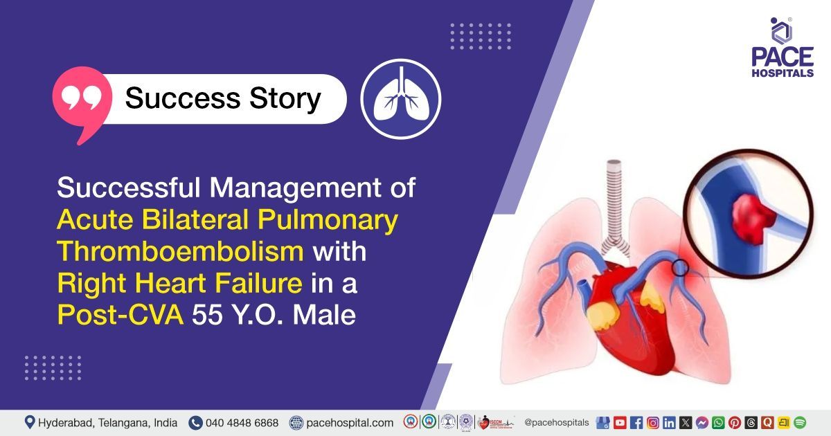
Acute Bilateral Pulmonary Thromboembolism with Respiratory Failure & Right Heart Failure in a Post-CVA Patient
PACE Hospitals
PACE Hospitals’ expert Pulmonology team successfully managed a 55-year-old male patient diagnosed with acute bilateral pulmonary thromboembolism (PTE) associated with acute respiratory failure and right heart failure in a patient with a previous cerebrovascular accident (CVA). The treatment focused on restoring breathing, improving oxygenation, preventing further clot progression, and stabilising heart and lung function through anticoagulation, oxygen therapy, nebulisation, and supportive critical care.
Chief Complaints
A 55-year-old male patient with a body mass index (BMI) of 19 presented to the Pulmonology Department at PACE Hospitals, Hitech City, Hyderabad, with complaints of shortness of breath, dryness of the throat, and cough with scanty expectoration for the past 15 days. He also reported worsening breathlessness over the preceding few months, with marked deterioration during the last 15 days.
Past Medical History
The patient was a known case of cerebrovascular accident (CVA) with right-sided hemiparesis and was on treatment. He had reportedly discontinued his regular medications before the onset of worsening symptoms. He had no known history of hypertension, diabetes mellitus, or tuberculosis. He was a former smoker who had quit smoking five years earlier.
On Examination
On examination, the patient was conscious, coherent, and oriented but was in severe respiratory distress. He required immediate oxygen support because of significant breathing difficulty. Lower limb swelling was noted on walking, while there was no wheezing, chest tightness, fever, weight loss, or loss of appetite. Considering the severity of his condition, he was admitted to the intensive care unit for close monitoring and further management.
Diagnosis
Upon admission to PACE Hospitals, following a detailed clinical assessment, the Pulmonology team evaluated the patient for severe shortness of breath, respiratory distress, dryness of the throat, cough with scanty expectoration, and a history of cerebrovascular accident with right-sided hemiparesis.
CT pulmonary angiography performed before referral revealed bilateral segmental and subsegmental pulmonary thromboembolism. Cardiac evaluation through 2D echocardiography showed dilatation of the right atrium and right ventricle, along with severe pulmonary arterial hypertension and severe tricuspid regurgitation, indicating significant strain on the right side of the heart.
Laboratory investigations showed elevated D-dimer and NT-proBNP levels, supporting the presence of acute pulmonary thromboembolism and cardiac stress. Doppler studies of both lower limbs and the neck veins did not reveal any significant venous thrombosis. The remaining blood investigations were largely within acceptable limits.
Based on the clinical presentation, CT pulmonary angiography, cardiac findings, and laboratory investigations, the patient was diagnosed with acute bilateral pulmonary thromboembolism associated with acute respiratory failure and right heart failure in a patient with a previous cerebrovascular accident.
Based on the confirmed diagnosis, the patient was advised to undergo Acute Pulmonary Thromboembolism Treatment in Hyderabad, India, under the expert care of the Pulmonology Department at PACE Hospitals.
Medical Decision-Making (MDM)
After a detailed consultation with the consultant interventional pulmonologist Dr. Pradeep Kiran Panchadi, and cross consultations with nephrologist Dr. A. Kishore Kumar, the neurophysician, ENT surgeon, and cardiologist, a comprehensive evaluation was performed to determine the most appropriate diagnostic and therapeutic approach.
Considering the patient’s history of cerebrovascular accident with right-sided hemiparesis, progressive breathlessness, severe respiratory distress, and clinical findings suggestive of pulmonary thromboembolism, further evaluation confirmed acute bilateral segmental and subsegmental pulmonary thromboembolism with acute respiratory failure and right heart failure. CT pulmonary angiography showed bilateral pulmonary emboli, while 2D echocardiography revealed right atrial and right ventricular dilatation, severe pulmonary arterial hypertension, and severe tricuspid regurgitation. Doppler studies of both lower limbs and neck veins were normal.
Based on the clinical assessment, imaging findings, and laboratory investigations, it was determined that intensive care management with oxygen support, low molecular weight anticoagulants, diuretics, nebulization, breathing exercises, and physiotherapy was the most appropriate and effective management strategy. This approach was chosen to improve oxygenation, prevent further clot formation, reduce strain on the right side of the heart, manage pedal edema, relieve respiratory distress, and support overall recovery.
The patient and his family members were counselled regarding the diagnosis, clinical findings, treatment plan, importance of anticoagulant compliance, regular coagulation tests, breathing exercises, physiotherapy, follow-up investigations, and continued review with the pulmonologist and cardiologist.
Treatment
Following the decision, the patient underwent treatment for acute bilateral pulmonary thromboembolism, acute respiratory failure, and right heart failure associated with cerebrovascular accident at PACE Hospitals, Hyderabad, under the expert supervision of the Pulmonology team.
During his hospital stay, the patient received anticoagulant therapy to prevent further clot formation and support the gradual resolution of the pulmonary embolism. Diuretic therapy was given to reduce pedal edema and relieve strain on the right side of the heart.
Nebulisation therapy, oxygen support, breathing exercises, incentive spirometry, physiotherapy for both lower limbs, nutritional support, and other supportive measures were provided to improve respiratory function and overall recovery.
The patient was closely monitored for oxygen saturation, respiratory rate, haemodynamic stability, neurological status, pedal edema, and any signs of bleeding or worsening breathlessness.
After clinical improvement and a reduction in oxygen requirement, the patient was shifted from the intensive care unit to the general ward and later discharged in a stable condition with advice.
Discharge Medications
At discharge, the patient was advised to continue medications to prevent the formation of new blood clots, reduce excess fluid accumulation and leg swelling, control cholesterol levels, relieve breathing difficulty, manage allergy-related throat irritation, protect the stomach from acidity, and prevent seizures related to the previous cerebrovascular accident.
Nebulisation was continued as advised to support easier breathing. The patient was also instructed to follow the prescribed dosage and duration carefully, undergo regular blood-clotting tests, and attend scheduled pulmonology and cardiology follow-up visits.
Advice on Discharge
The patient was advised to take all prescribed medications regularly, continue breathing exercises, incentive spirometry, and lower-limb physiotherapy, and avoid prolonged immobility. A protein-rich diet, adequate hydration, and gradual physical activity were also recommended.
Emergency Care
The patient was informed to contact the emergency ward at PACE Hospitals in case of sudden or increasing breathlessness, chest pain, reduced oxygen saturation, coughing up blood, fainting, confusion, bluish discoloration of the lips or fingers, increased leg swelling, unusual bleeding, or any other alarming symptoms.
Review and Follow-up Notes
The patient was advised to return for a follow-up consultation with the Pulmonologist in Hyderabad at PACE Hospitals, after 1 month.
Conclusion
This case highlights the successful multidisciplinary management of acute bilateral pulmonary thromboembolism complicated by acute respiratory failure and right heart failure in a patient with a previous cerebrovascular accident. Early diagnosis, prompt intensive care, timely anticoagulation, and coordinated supportive treatment resulted in significant clinical improvement and a stable recovery at discharge.
Importance of Early Diagnosis and Treatment of Acute Pulmonary Thromboembolism
Acute pulmonary thromboembolism is a medical emergency that requires rapid diagnosis and prompt treatment to prevent life-threatening complications such as respiratory failure and right heart failure. A pulmonologist or pulmonology doctor usually assesses the patient through clinical evaluation, CT pulmonary angiography, laboratory investigations, and cardiac assessment. Early anticoagulation, oxygen therapy, and supportive care help improve blood flow through the lungs, support oxygen delivery, and reduce strain on the heart. A multidisciplinary approach involving a pulmonologist or pulmonology doctor, cardiologists, neurologists, and other specialists may be required in complex cases. Timely treatment, close monitoring, and regular follow-up can improve recovery and reduce the risk of recurrent pulmonary embolism.
Frequently Asked Questions (FAQs)
Why was intensive care admission required for this patient with pulmonary thromboembolism?
Intensive care was required because the pulmonary embolism had caused severe breathing difficulty, acute respiratory failure, and strain on the right side of the heart. ICU care allowed continuous monitoring of oxygen levels, breathing, blood pressure, heart function, and neurological condition. Immediate oxygen support and anticoagulation could also be provided safely.
How does pulmonary thromboembolism lead to right heart failure?
Pulmonary thromboembolism blocks the normal flow of blood through the arteries in the lungs. As a result, the right side of the heart has to work harder to push blood forward. If the pressure becomes too high, the right ventricle may become enlarged and weak, which can lead to right heart failure.
Why was CT pulmonary angiography important in this case?
CT pulmonary angiography helped identify blood clots within the segmental and subsegmental arteries of both lungs. It also provided information about the location and extent of the blockage. Imaging is necessary because pulmonary embolism can resemble pneumonia, heart disease, and other respiratory conditions.
What did the enlarged right atrium and right ventricle indicate?
Enlargement of the right atrium and right ventricle suggested that the blood clots were placing significant pressure on the right side of the heart. These findings, together with severe pulmonary hypertension and tricuspid regurgitation, indicated right heart strain. Such patients require close cardiac and respiratory monitoring.
Why were anticoagulants given even though the leg Doppler study was normal?
A normal leg Doppler does not rule out pulmonary embolism. The original clot may have already travelled to the lungs, may have resolved in the leg, or may have formed in an area not detected during the study. Since CT pulmonary angiography confirmed pulmonary embolism, anticoagulation was required to prevent existing clots from enlarging and new clots from forming.
Why was the previous cerebrovascular accident considered before starting treatment?
A history of cerebrovascular accident is important because blood-thinning treatment can increase the risk of bleeding. The medical team must consider the type and timing of the previous stroke, current neurological condition, ongoing medications, and overall bleeding risk. Specialist consultation helps determine the safest treatment approach.
How do anticoagulants help in pulmonary thromboembolism?
Anticoagulants do not immediately dissolve the existing clot. They prevent the clot from becoming larger and reduce the formation of additional clots while the body gradually breaks down the blockage. They are the main treatment for most patients with confirmed acute pulmonary embolism.
Why were oxygen therapy, nebulisation, and diuretics required?
Oxygen support was given to help the patient breathe comfortably and maintain proper oxygen levels. Nebulisation was used to ease breathing, while diuretics helped reduce fluid buildup and swelling in the legs. This also helped lessen the strain on the right side of the heart.
How long does recovery from acute pulmonary thromboembolism take?
Recovery differs according to the size of the clot, severity of heart strain, previous health conditions, and response to treatment. Breathlessness and tiredness may improve gradually over several weeks or months. Many patients recover well when pulmonary embolism is diagnosed early and anticoagulation is taken exactly as prescribed.
How can another pulmonary embolism be prevented after discharge?
Another pulmonary embolism can be prevented by taking the prescribed blood-thinning treatment regularly and not stopping it without medical advice. Staying active, performing lower-limb exercises, drinking enough fluids, avoiding smoking, and not sitting or lying down for long periods can also lower the risk of new clots. Sudden breathlessness, chest pain, coughing up blood, fainting, or worsening leg swelling should be treated as an emergency.
Share on
Request an appointment
Fill in the appointment form or call us instantly to book a confirmed appointment with our super specialist at 04048486868
Appointment request - health articles
Recent Articles