Pace Hospitals | Best Hospitals in Hyderabad, Telangana, India

Blog Post

Breast Self Examination - Steps, Purpose, Importance and Advantages

Pace Hospitals

Breast Self Examination Introduction

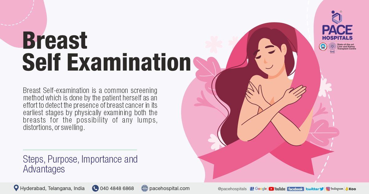
Breast Self-examination is a common screening method which is done by the patient herself as an effort to detect the presence of breast cancer in its earliest stages by physically examining both the breasts for the possibility of any lumps, distortions, or swelling.

Necessity and Purpose of Breast Self Examination

A regular breast cancer screening is a necessary as early detection is a major factor in treating cancer. Cancers diagnosed at an earlier stage have a better prognosis and are easier to treat. Reducing the likelihood of dying from breast cancer requires regular screening.


Breast self-examination as the most economical and potential screening procedure which had provided significant results. Women are encouraged to get to know their breasts and become more attuned to any changes, without resorting to a strict regimen of self-examinations at predetermined intervals. Self-examination of the breast was very much helpful in low-resource countries where access to mammography is limited.

Breast Self Examination Test

A free-standing or wall-mounted mirror is all that is required to conduct a thorough breast self-examination through visual inspection. In case the patient is lying on their back to complete the procedure, a pillow may be used to prop up the back or shoulder. 

Breast Self Examination Steps

A breast self-examination is a step-by-step procedure where all females can use to examine their breasts. These are the steps for breast self-examination that every woman should follow:

  • Female should remove any kind of garment on her upper body before attempting for a breast self-examination.
  • Female must be in front of a large mirror which reflects at least her upper body during the breast self-examination.
  • Breast self-examination starts with the visual inspection of the breasts in the reflection. 
  • While typically one breast may be larger than the other, new disparities in size should be noted such as dimpling in the skin, changes in breast shape, swelling in both the breasts, any rashes, erythema, puckering, textural anomalies resembling an orange peel (peau d’orange).
  • Any changes in the nipples must be monitored for any scaling, erythema, pruritis, oedema, discharge, or any new inversion.
  • Asymmetric venous distribution or dilation should also be taken into consideration.
  • The visual survey of the breast tissue requires an inspection from all the three angles,
  1. with arms at the side,
  2. arms raised above the head while bending forward, and
  3. hunched over with the hands placed on the hips. 


These steps are done to flex the chest muscles so that if any abnormalities present will appear. 

Each of these positions should be observed in a mirror from three views:

  1. a direct view, 
  2. right profile, 
  3. left profile.


Once the visual examination is completed, physical (tactile) examinations must be commenced, in which a self-assessment will be made by feeing the chest area by the female. 

  • The female should roll onto her left side and rest her right-hand palm up on her forehead in order to facilitate a right breast examination. This manoeuvre improves positioning. 
  • When feeling for different layers of breast tissue, it's best to use the pad of the middle finger to make small, medium, and large circles at different pressures. 


The medial half of the breast is gently pressed for any abnormalities from the armpit to the nipple, and the vertical half is palpated (gently pressed) from the clavicle bone to just below the bra line.


It's important to keep the fingers in constant contact with the skin as they move across the breasts so as not to miss any tissue planes.

  • To evaluate the underside of the breasts, the female must assume a supine position (lying on her back). The right-hand must be taken off the forehead, and placed at a right angle on the examination surface.
  • The inner breast, including the nipple and sternum, is palpated in the same way. 


Consistent and repeatable results require a methodical, systematic approach to evaluation. 

The visual and physical examination of the left breast entails the same positioning of the patient and the same series of manoeuvres as those used for the examination of the right breast. 


The female must remain calm in case if the female finds any kind of lump or any other bothersome changes in either or both the breasts. It must be understood that most of the findings from the self-examination of the breast typically are not signs of breast cancer. The patient should consult the doctor at once if any of the above explained abnormalities are found during the breast self-examination. 

breast self examination steps | breast self examination poster | breast self examination health education | breast lump self examination

Indication of breast self-examination 

Although breast self-examination was indicated for the self-awareness of women for the early detection of breast cancer (by finding the lumps) during the days before the advent of mammography; it also helped in the early detection of various other conditions such as; 

  • Phyllodes tumour (rare breast tumours starting in the connective tissue of the breast, rather than ducts or glands)
  • Fibroadenoma (benign tumours of fibrous and glandular tissue which are solid and usually move quite easily)
  • Cysts (build-up of fluid and can be round or oval and feel soft or hard)
  • Lipoma (harmless growth of fatty tissue)
  • Duct ectasia (benign (non-cancerous) breast condition of widening of milk duct and thickening of its walls)
  • Sebaceous cyst (cysts from sebaceous glands. Sebaceous glands are found underneath the skin secreting sebum. Sebaceous cyst can develop if the gland or its duct gets damaged or blocked)
  • Galactocele (benign breast lesions commonly seen in young lactating women which occur due to the blockage of the ducts due to the stagnant milk after lactation)
  • Fat necrosis (death of fat tissue due to injury and loss of blood supply)
  • Tuberculous abscess (accumulation of pus due to tuberculous)

Advantages of breast self-examination

Breast self-examinations can help in the early detection of breast cancer early, thereby improving the chances of a full recovery. Overall five-year breast cancer survival rate is 89.7%. Early diagnosis improves long-term outcomes.


Five-year survival rate for women diagnosed at 

  • Stage 0 or Stage 1, the survival rate is 98.8% or 
  • Stage 2, the survival rate is 93%. 
  • Stage 3, the survival rate is 72% 
  • Stage 4, the five-year survival rate is 22%.

Importance of breast self-examination

The importance of breast self-examination was noticed when an increased proportion of women with early diagnosis of breast cancer were treated in time successfully.


When the diagnostic procedure of breast cancer was introduced, a small fraction of medical fraternity listed the potential burden of false-positive results associated with pointless imaging tests and unnecessary biopsies which resulted in the resistance against breast self-examination.


Resistance was soon abandoned due to the 

  • high incidence of breast cancer, 
  • positive anecdotal experiences reported by patients and practitioners, and 
  • intention to empower women through self-diagnosis.


Recognizing the importance of early breast cancer detection through breast self-examination, several organisations promoted the breast self-examination as a potential screening procedure, encouraging women to be cognizant of any changes in their body 

Contraindications of breast self-examination

There are no absolute contraindications or risk factors in the implementation of a breast self-examination programme. Finding a lump in your breast can be alarming, but it must be understood that most breast lumps aren't malignant (cancerous). They are caused by other benign conditions.

Where are breast lumps usually found?

If each of the breasts can be divided into 4 quadrants, then it can be said that the upper outer quadrant area, which is nearest to the armpit, could be the most common area to find lumps. 

breast cancer self examination images | breasts quadrants | objective of breast self examination | need of breast self examination

Frequently asked questions

  • How to self-examine for breast cancer?

    Self-examination for breast cancer can be done by the patient herself. By physically examining both the breasts for the possibility of any lumps, distortions, or swellings, the early detection of breast cancer can be achieved. A large mirror can be helpful for effective self-examination of breast cancer.

  • How frequently should self-examination of breast be done?

    The best time for self-examination for breast cancer is a few days after the menstrual cycle each month. Since any swollenness or tenderness of the breast with the menstrual cycle is eliminated within a few days after the menstrual cycle, this time could be perfect for the self-examination of breast cancer.


    A diary can be maintained by the patient to record the self-examination for breast cancer which could be helpful for future diagnosis by the oncologist.


    With regular home breast exams, the chances of early detection of breast cancer increase thus enabling a quicker doctor consultation. 

  • How much time needed for completing breast self-examination?

    Since a breast self-exam takes only a few minutes, it can be done during the non-productive tasks of the daily routines such as: 

    • Dressing or undressing 
    • Lying in bed in the morning or at bedtime.
    • Taking a shower.
  • Why we do breast self-examination?

    With regular home breast exams, the chances of early detection of breast cancer in women increase thus enabling a quicker doctor consultation. Breast cancer is the most prevalent cancer in the world and early detection is one of the best ways to prevent it.

  • Can a breast lump grow fast?

    There are various factors for the growth of tumour. A 2008 study demonstrated that

    • 5% of tumours grew from 10-20 mm in less than 1.2 months, and 
    • 5% took more than 6.3 years. 
    • The mean time a tumour needed to grow from 10-20 mm in diameter was estimated as 1.7 years, increasing with age.
  • Why we are discussing over breast self-examination?

    Breast cancer is the most prevalent cancer in the world and early detection is one of the best ways to prevent it. Self-examination of the breast is the cheapest diagnostic procedure the patient can cater to without any loss of her modesty. With regular home breast exams, the chances of early detection of breast cancer increase thus enabling a quicker doctor consultation.

  • What does a cancerous lump feel like?

    A cancerous lump could be tender, soft rounded, and in few cases, it is also painful which occurs anywhere in the breast. Feeling breast lumps or changes in the texture is a bit difficult in case of women with dense, fibrous breast tissue. 

  • How do you know if a lump is moveable?

    A doctor consultation for examination must be sought in the discovery of any abnormality in the breasts - even movable or immovable lumps discovered from self-examination. Lumps that can easily move with the pads of your fingers are not usually cancerous but could be of the following ailments: 

    • Fibroadenomas (benign tumours of fibrous and glandular tissue which are solid and usually move quite easily)
    • Cysts (build-up of fluid and can be round or oval and feel soft or hard)
    • Papilloma (wart-like lumps developing in the lining of mammary ducts, usually near the nipple)
    • Breast abscess (accumulation of pus due to infection)
    • Necrosis (death fatty tissues in the breast due to an injury to breasts. Necrosis is not cancer)
    • Sclerosing adenosis (overgrowth of tissue in breast lobules and can be mistaken for breast cancer)
    • Lipoma (harmless growth of fatty tissue)
  • What percentage of breast lumps are cancerous?

    The majority of the breast lumps – around 80% of the breast lumps which underwent biopsy are benign (non-cancerous). Even if the lump is found to be cancer, since it would be found in an early cancer detection stage, early treatment can stop cancer. 

  • How long should you wait to get a breast lump checked?

    A doctor consultation must be necessary to examine the development of breast lumps especially 

    • If the lump is firm or fixed
    • If the presence of a lump is seen even after four to six weeks
    • If there are any changes seen on the skin of the breast, such as redness, crusting, dimpling or puckering
    • If there is a bloody discharge, possibly from the nipple
    • If the nipple is turned inward and isn't normally positioned that way
    • If there is a lump in the armpit which gets bigger

Request an appointment

Fill in the appointment form or call us instantly to book a confirmed appointment with our super specialist at 04048486868

Appointment request - health articles

World Oral Health Day, 20 March 2025 | Theme, Importance & History | Theme of World Oral Health Day
By Pace Hospitals March 15, 2025
World Oral Health Day is a global healthcare event with this year's (2025) theme "A HAPPY MOUTH IS… A HAPPY BODY" which is observed on the 20th of March to raise awareness about oral hygiene.
National Immunization Day or National Vaccination Day, 16 March 2025 – Theme & Importance
By Pace Hospitals March 13, 2025
National Immunization Day (also called National Vaccination Day) is a nationwide healthcare event, typically observed on the 16th of March every year, intending to encourage the deserved recognition of the policymakers, the healthcare workers as well as the other stakeholders and their work in conducting several successful vaccination drives.
PACE Hospitals Podcast with Dr K Meena on lymphoma Types, Causes, Symptoms & Treatment
By Pace Hospitals March 13, 2025
Tune in to the PACE Hospitals Podcast with Dr K Meena to explore lymphoma, its symptoms, causes, risk factors, diagnosis, and advanced treatment options, including chemotherapy, immunotherapy, and targeted therapy.
Case study of a 34-Y/O woman suffering from gallstone pain successfully treated at PACE Hospitals
By Pace Hospitals March 13, 2025
Explore the case study of a 34-year-old woman suffering from gallstone pain who underwent a successful laparoscopic cholecystectomy at PACE Hospitals, Hyderabad, ensuring a quick recovery and restoring her health and quality of life.
cardiac arrest symptoms | reasons for cardiac arrest | cardiac arrest treatment in India
By Pace Hospitals March 13, 2025
Cardiac arrest occurs when the heart stops pumping blood to vital organs, leading to unconsciousness within seconds. Learn about its symptoms, causes, types, risk factors, diagnosis, complications, treatment, and prevention.
Acute Necrotizing Pancreatitis symptoms  | necrotizing pancreatitis treatment in India
By Pace Hospitals March 13, 2025
Necrotizing pancreatitis is a severe form of pancreatitis that leads to pancreatic tissue death. Learn about its symptoms, causes, risk factors, complications, diagnosis, treatment, and prevention.
Case Study: Total Knee Replacement Surgery for a 51-Year-Old Patient at PACE Hospitals
By Pace Hospitals March 12, 2025
Explore the case study of a 51-year-old patient who underwent a successful total knee replacement surgery performed by the Orthopaedics team at PACE Hospitals. This procedure not only alleviated her severe knee pain but also restored her mobility and greatly enhancing her quality of life.
World Kidney Day 13 March 2025 | Theme and Importance
By Pace Hospitals March 10, 2025
World Kidney Day is a global healthcare event celebrated on the 2nd Thursday in March every year since 2006. The epidemic of chronic kidney disease in Indians is seen with a scope of increasing diabetes load, hypertension, and an aging population; it will only worsen.
Case study of a 63-Y/O male who suffered from GERD and hiatal hernia treated at PACE Hospitals
By Pace Hospitals March 8, 2025
Explore the case study of a 63-year-old male who suffered from GERD and hiatal hernia. After undergoing advanced laparoscopic fundoplication at PACE Hospitals, he found lasting relief and improved his quality of life.
Show More

Share by: