
Successful eTEP-TAR Repair of Incisional and Umbilical Hernias in a 52-Year-Old Patient
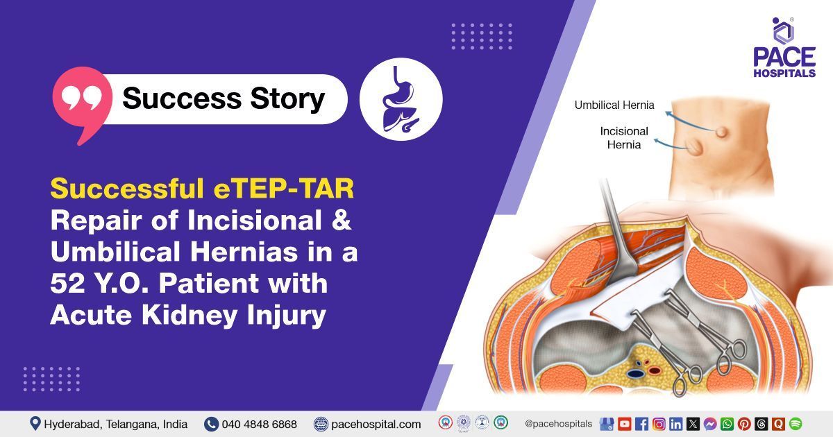
The Surgical Gastroenterology team at PACE Hospitals successfully performed an Extended Totally Extraperitoneal Repair (eTEP) with a Right Transverse Abdominis Release (TAR) on a 52-year-old male who had been struggling with a right subcostal incisional hernia and an umbilical hernia, aiming to improve recovery and minimize complications.
Chief Complaints
A 52-year-old male patient presented to the Gastroenterology Department at
PACE Hospitals, Hitech City, Hyderabad, with complaints of swelling near the right subcostal region (located in the right upper quadrant of the abdomen). In addition, the patient had a concurrent
umbilical hernia, further complicating his abdominal condition. The swelling in the subcostal region had progressively worsened over time, causing increasing discomfort.
Medical History
The patient had a known medical history of an open cholecystectomy performed in 2012 to remove the gallbladder, which was necessitated by complications such as gallstones or gallbladder inflammation. The open cholecystectomy involved making a larger abdominal incision to access and remove the gallbladder, a procedure typically chosen when laparoscopic methods were not feasible due to complications or the patient's specific condition. This surgical history was relevant to his current presentation, as the previous incision site likely contributed to the development of the right subcostal incisional hernia.
Right subcostal incisional hernia occurs at the site of a previous surgical incision, often due to abdominal wall weakness. An umbilical hernia involves protrusion of abdominal tissue through the abdominal wall at the belly button, which is common in both infants and adults. Both types may require surgical intervention to prevent complications like bowel obstruction or strangulation.
Diagnosis
Upon admission to PACE Hospitals, the patient's vital signs were stable, showing no immediate signs of distress. Following a thorough physical examination, the doctors identified swelling in the right subcostal region (in the right upper quadrant of the abdomen) along with the presence of an umbilical hernia.
Diagnostic tests, including ultrasound imaging and CT scans, later confirmed the diagnosis of a right subcostal incisional hernia and a concurrent umbilical hernia. The clinical findings, in combination with the patient's surgical history of an open cholecystectomy, helped establish the basis for the diagnosis and subsequent treatment plan.
Medical Decision Making (MDM)
After consulting with the interventional and consultant gastroenterologists, Dr. Suresh Kumar S, along with other consultants such as Dr. A Kishore Kumar, conducted a comprehensive evaluation was conducted to determine the most appropriate course of treatment for the patient.
Their collective expertise, drawing on years of experience in abdominal surgery and hernia repair, led to the conclusion that an Extended Totally Extraperitoneal Repair (eTEP) in combination with Right Transverse Abdominis Release (TAR) would be the most appropriate treatment for the patient's right subcostal incisional hernia and umbilical hernia. The decision was based on the need for a minimally invasive yet effective approach to managing both hernias while minimizing post-operative complications and promoting optimal recovery.
Surgical procedure
Upon admission, the patient was thoroughly evaluated and deemed fit for surgery. Following this assessment, the patient underwent an Extended Totally Extraperitoneal Repair (eTEP) in combination with Right Transverse Abdominis Release (TAR).
The procedure was planned carefully to address both hernias. During the procedure, the Extended Totally Extraperitoneal (eTEP) Repair, combined with a Right Transverse Abdominis Release, was chosen as the surgical method due to the complexity of the patient's right subcostal incisional hernia and umbilical hernia. This minimally invasive technique involved making small incisions, through which a laparoscope was inserted to repair the hernia without entering the peritoneal cavity. The Right Transverse Abdominis Release (TAR) procedure involved cutting the muscle layer (specifically the transverse abdominis muscle) for enhanced exposure and access to the hernia site.
During the surgery, mesh was used to reinforce the abdominal wall, ensuring a robust repair. The decision to combine these techniques was based on their ability to improve surgical outcomes by reducing recurrence rates and accelerating recovery time compared to traditional open surgery. This innovative approach was selected to provide the patient with the best possible results, minimizing potential complications and ensuring a faster return to normal activities.
Findings
During the procedure, the surgical team successfully accessed the eTEP plane, which is located posterior to the rectus muscle and anterior to the posterior rectus sheath, starting from above.
Dense intra-abdominal adhesions were observed near the previous cholecystectomy site, likely a result of the patient’s prior gallbladder removal.
A 6x6 cm hernial defect was found, with the omentum (fatty tissue) protruding through the defect Additionally, multiple small hernia defects were identified along the right subcostal and anterior abdominal wall, also containing omentum.
The umbilical hernia was successfully closed using V-lock no. 1 sutures. A 45x45 cm Dynamesh was placed to reinforce the abdominal wall.
However, complete mesh approximation could not be achieved, so a small midline incision was made to allow for proper buttressing of the mesh, ensuring a secure repair. This approach helped provide optimal support to the abdominal wall, reducing the risk of recurrence.
Postoperative Care
The procedure was smooth and without complications. On post-operative day 1 (POD-1), the patient was started on a liquid diet, which was gradually advanced to a soft diet as tolerated. However, during the recovery phase, the patient's creatinine levels were found to be elevated, along with the presence of proteinuria, which raised suspicion for drug-induced acute kidney injury (AKI) or an underlying kidney condition.
To investigate the renal concerns further, relevant investigations were conducted, including serum creatinine tests, urinalysis, and renal function tests. Based on the results, the patient was managed conservatively, with close monitoring of kidney function, ensuring appropriate treatment was provided to address any renal issues. This included adjusting medications and fluids as needed while ensuring optimal recovery from both the hernia surgery and renal concerns.
Discharge notes
The patient had a satisfactory postoperative recovery and was discharged in a hemodynamically stable condition. Before discharge, the patient received the discharge instructions.
Discharge Medications
Upon discharge, the patient was prescribed antibiotics, proton pump inhibitors (PPIs), pain relievers, laxatives, anticoagulants, mineral supplements, and opioids with instructions to continue a normal diet.
Advice on Discharge
The patient was advised to wear an abdominal binder for 6 weeks, avoid strenuous activities, and continue taking prescribed medications as directed.
Emergency Care
The patient’s guardians were informed to seek admission to the Emergency Ward of PACE Hospitals, if they observe any concerning symptoms, including fever, abdominal pain, or vomiting. These signs could indicate potential complications, such as infection, intestinal obstruction, or other post-operative issues, and Immediate medical attention would be necessary to address them promptly.
Review and follow-up notes
The patient was advised to follow up with the nephrologist, Dr. Kishore, after 1 week, along with the fundus examination report. Additionally, a follow-up appointment was scheduled at the Surgical Gastroenterology OPD after 1 week.
Acute Kidney Injury (AKI) Management Following Hernia Repair
Acute Kidney Injury (AKI) can occur during hernia repair, especially in cases involving complex hernias or large ventral hernias, due to factors such as increased intra-abdominal pressure, reduced kidney perfusion, and potential complications arising from surgery, underlying conditions, medications, or fluid imbalances. Additionally, pressure on surrounding abdominal structures during the procedure can exacerbate these issues.
As a result, the patient may experience reduced renal perfusion, elevated creatinine levels, proteinuria, and decreased urine output. The presence of certain hernias, particularly those involving the right subcostal and umbilical regions, complicates the management of AKI, as abdominal pressure and discomfort can hinder optimal kidney function during recovery.
In these cases, close monitoring of renal function, conservative management (including hydration, medication adjustments, and addressing fluid imbalances), and further diagnostic evaluation are crucial to prevent the progression of AKI to severe renal impairment. Early intervention, timely adjustments, and a multidisciplinary approach are essential to mitigate risks and ensure the best possible outcomes for patients undergoing hernia repair surgery.
Share on
Request an appointment
Fill in the appointment form or call us instantly to book a confirmed appointment with our super specialist at 04048486868
Appointment request - health articles


Our Locations – Find the Best Hospital Near You
Metro Pillar Number C1772, Beside Avasa Hotel, Hitech City Road, Near HITEC City Metro Station, Hyderabad, Telangana, India.
Mythri Nagar, Beside South India Shopping Mall, Hafeezpet, Madeenaguda, Hyderabad, Telangana, India.
040 4848 6868
Payment in advance for treatment at PACE Hospitals, Hyderabad, Telangana, India (Pay in INR ₹)
For Bank Transfer:-
- Bank Name: HDFC
Company Name: Pace Hospitals
A/c No.50200028705218
IFSC Code: HDFC0000545 - Bank Name: STATE BANK OF INDIA
Company Name: Pace Hospitals
A/c No.62206858997
IFSC Code: SBIN0020299
Scan QR Code by Any Payment App (GPay, Paytm, Phonepe, BHIM, Bank Apps, Amazon, Airtel, Truecaller, Idea, Whatsapp etc).

CONTACT US
Call: +914048486868
WhatsApp: +918977889778
Email: info@pacehospitals.in
FOLLOW US
SUBSCRIBE
Subscribe to our newsletter and stay updated with the latest health information.
ABOUT US
QUICK LINKS
Disclaimer
General information on healthcare issues is made available by PACE Hospitals through this website (www.pacehospital.com), as well as its other websites and branded social media pages. The text, videos, illustrations, photographs, quoted information, and other materials found on these websites (here by collectively referred to as "Content") are offered for informational purposes only and is neither exhaustive nor complete. Prior to forming a decision in regard to your health, consult your doctor or any another healthcare professional. PACE Hospitals does not have an obligation to update or modify the "Content" or to explain or resolve any inconsistencies therein.
The "Content" from the website of PACE Hospitals or from its branded social media pages might include any adult explicit "Content" which is deemed exclusively medical or health-related and not otherwise. Publishing material or making references to specific sources, such as to any particular therapies, goods, drugs, practises, doctors, nurses, other healthcare professionals, diagnoses or procedures is done purely for informational purposes and does not reflect any endorsement by PACE Hospitals – your trusted hospital near me.

