Pancreatic Cysts - Types, Causes, Symptoms, Complications, Treatment & Prevention
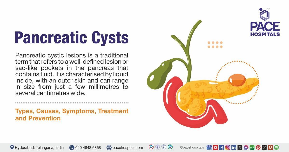
Pancreatic cystic lesions are a traditional term that refers to a well-defined lesion or sac-like pockets in the pancreas that contains fluid.
They are characterised by liquid inside, with an outer skin, and can range in size from just a few millimetres to several centimetres wide. However, the majority are small and less than a centimetre or two in diameter.
Prevalence of pancreatic cyst
The prevalence of pancreatic cysts varies greatly depending on the imaging method used, ranging from 0.21% with ultrasound to 50% in autopsy studies, and it increases with age.
Globally, pancreatic cystic lesions (PCLs) are common, with multiple studies suggesting a prevalence ranging from 2% to 38% depending on the imaging modality used, with an overall prevalence of around 15%.
While data on the prevalence of pancreatic cysts in India is limited, studies suggest a prevalence of around 2.5% in asymptomatic populations, increasing with age. However, some studies show higher rates, particularly in older age groups.

Types of Pancreatic Cysts
Pancreatic cystic lesions can be classified as:
Benign cysts
non-cancerous, e.g., simple cysts, Pancreatic pseudocysts, and serous cystic neoplasms (SCNs).
Cysts with malignant potential
e.g., mucinous cystic neoplasms (MCNs) and intraductal papillary mucinous neoplasms (IPMNs).
Malignant cysts
Neoplastic cysts such as pancreatic adenocarcinomas with cystic degeneration and cystic pancreatic neuroendocrine tumors.
Pseudocysts: Pseudocysts are the most common type, which emerges following either acute or chronic pancreatitis. They usually manifest as one or more unilocular cysts that may contain debris and cannot become malignant (not able to develop into cancer). Intervention is only necessary for symptomatic pseudocysts; the majority resolve on their own.
Serous cystadenomas: Serous cystadenomas are slow-growing, benign lesions that primarily affect women between the ages of five and seven. Although they might appear as solid, macrocystic, or unilocular lesions, these cysts typically have a microcystic (honeycomb) look. They are incapable of developing into cancer.
Mucinous Cystic Neoplasm: The less prevalent mucinous cysts are called MCNs and are typically seen in the distal pancreas. They are single, thick-walled, primarily unilocular cysts.
They primarily affect women between the ages of 40 and 50 and can develop into cancer. Furthermore, they characteristically contain ovarian-like stroma and almost exclusively affect women in the fourth to sixth decades of life.
Intraductal Papillary Mucinous Neoplasm: IPMNs are the most common type of mucinous cystic lesions. Pancreatitis may coexist with intraductal papillary mucinous neoplasm (IPMNs); men are more likely than women to have this condition. These ductal cell-derived neoplasms are frequently multifocal and spread throughout the pancreas. IPMNs have the potential to become malignant.
Solid Pseudopapillary Neoplasm: The rarest pancreatic cyst is a solid pseudopapillary neoplasm, primarily affecting women in their second or third decade. These well-defined, heterogeneous lesions, which can be found all over the pancreas, contain solid and cystic components and occasionally irregular calcifications. Although most pancreatic cysts are benign, some have the potential to become cancerous.
Revised WHO histological classification of pancreatic cystic neoplasms (WHO pancreatic cyst classification):
- Serous cystic neoplasm (SCN)
- Serous cystadenoma
- Serous microcystic adenoma (Benign)
- Serous oligocystic adenoma (Benign)
- Serous cystadenocarcinoma (Malignant)
- Mucinous cystic neoplasm (MCN)
- Mucinous cystadenoma (Benign)
- Mucinous cystic neoplasm
- Low-grade (Borderline)
- High-grade (Carcinoma in situ)
- Mucinous cystadenocarcinoma
- Non-invasive (Malignant)
- Invasive (Malignant)
- Intraductal papillary mucinous neoplasm (IPMN)
- Intraductal papillary mucinous neoplasm
- Low-grade (Borderline)
- High-grade (Carcinoma in situ)
- Intraductal papillary mucinous carcinoma
- Non-invasive (Malignant)
- Invasive (Malignant)
- Solid pseudopapillary neoplasm
- Solid pseudopapillary neoplasm (Borderline)
- Solid pseudopapillary carcinoma (Malignant)
Pancreatic Cyst Symptoms
Most pancreatic cysts are not symptomatic (asymptomatic) and are discovered incidentally on diagnostic imaging that is carried out for an unrelated symptom or reason. The following are the common signs and symptoms of pancreatic cysts that can be seen in some patients, including:
- Jaundice: It is a rare symptom in patients with pancreatic cysts (less than 1%). However, it is a more common feature if cysts are huge or contain cancer cells. It happens due to a build-up of bilirubin (a chemical constituent of bile) in the blood. Jaundice can occur if a cyst in the pancreatic head compresses or blocks the ducts carrying bile from the liver.
- Abdominal pain: Small cysts, e.g. less than 2cm, usually do not cause pain. However, as cysts enlarge, they may exert pressure on the surrounding tissues and nerves, leading to persistent back or upper abdominal pain.
- Nausea and vomiting: Compression of the duodenum delays stomach emptying (gastric emptying), resulting in a feeling of fullness, which may lead to nausea and vomiting.
- Weight loss: It is a rare symptom; if the cysts are large, they can decrease the pancreas's ability to produce digestive enzymes, causing poor digestion of food and resulting in weight loss.
Potential symptoms related to specific types of cysts include:
Serous cystic neoplasm (SCN)
- Signs: Palpable mass
- Symptoms: Mostly asymptomatic
- If these SCNs are large, they cause gastric outlet obstruction and bile duct obstruction
Solid pseudopapillary neoplasm (SPN)
- Common symptoms: May present with nausea, abdominal pain, vomiting, and weight loss
- Other signs and symptoms: Anaemia, jaundice, gastric outlet or intestinal obstruction and pancreatitis.
Mucinous cystic neoplasm (MCN)
- Symptoms: Most patients are asymptomatic; possible symptoms include back pain, abdominal pain
- Signs: It may present as a palpable mass
- Other features: Recurrent pancreatitis, gastric outlet obstruction, jaundice, and weight loss are more familiar with malignant lesions.
Intraductal papillary mucinous neoplasm (IPMN)
- Symptoms and signs: Mostly asymptomatic, some patients have symptoms suggestive of chronic pancreatitis, which results from intermittent obstruction of the pancreatic duct with mucus plugs. Manifestations such as back pain, jaundice, weight loss, anorexia, steatorrhea, and diabetes are precursors of malignancy.
- Other features: Some patients have a long-standing history of recurrent acute pancreatitis
Neuroendocrine tumors (NET)
- May rarely manifested as cystic lesions
- Most are asymptomatic, but symptoms or signs due to the production of hormones may be present.

Pancreatic Cyst Causes
The exact reason for most pancreatic cysts is still idiopathic (unknown). However, some cysts might be partly hereditary and partly triggered by other factors, including:
- Genetic Disorders: As mentioned above, hereditary syndromes, including von Hippel-Lindau disease, cystic fibrosis, and polycystic kidney disease, are associated with pancreatic cysts.
- Pancreatitis: Pancreatitis, both acute and chronic, is a leading cause of pancreatic cysts. Inflammation can lead to the leakage of pancreatic enzymes, forming pseudocysts.
- Alcohol use: Over-alcohol consumption is the leading contributor to many health conditions, particularly pancreatitis, raising the risk for pancreatic cysts.
- Advancing age: Pancreatic cysts become more common as you get older. They affect an estimated 25% of people in their 70s.
- Family history: Some persons may be more likely to have pancreatic cysts if they have a family history of pancreatic cancer or if they have a hereditary syndrome associated with cysts.
- High Blood Calcium or Cholesterol Levels: Elevated calcium or fat levels in the blood can harm the pancreas and contribute to cyst formation.
- Infections: Rare infections like
tuberculosis or parasitic infections (e.g., tapeworms) can also trigger pancreatic cyst formation.
Pancreatic Cyst Risk Factors
Pseudocysts almost always occur following a bout of acute pancreatitis, a painful condition in which digestive enzymes become prematurely active and irritate the pancreas. Common risk factors for pancreatitis include:
- Heavy alcohol use: As mentioned above, heavy consumption of alcohol can increase the risk of pancreatitis, contributing to the significant risk of the formation of cysts in the pancreas.
- Gallstones: Gallstones can increase the risk of pancreatic cysts, particularly pancreatic pseudocysts, by causing pancreatitis by leaking the digestive enzymes, damaging the tissue, and potentially leading to cyst formation.
- Age: The risk of developing many pancreatic cysts, e.g. IPMN, increases with age. Some cysts are seen almost exclusively in women, like mucinous cystic neoplasms (more than 90%) and serous cystadenoma (more than 70%).

Pancreatic Cyst Complications
Complications are not common with pancreatic cysts, though they may include:
- Pancreatic cancer: Less than 1% of cysts can turn into cancer, most commonly adenocarcinoma, having the capability to spread. Hence, early detection is vital.
- Bile duct obstruction: A large cyst can obstruct the bile duct, causing jaundice (yellowing of the skin and eyes) and other biliary complications.
- Pancreatic Duct Obstruction: Certain mucinous cysts can obstruct the pancreatic duct, leading to problems with pancreatic function.
- Haemorrhage: Bleeding or cyst haemorrhage may occur within the cyst or surrounding tissues, resulting in internal bleeding and shock.
- Rupture: A ruptured cyst can cause internal bleeding and infection of the abdominal cavity (peritonitis), which can be life-threatening.
- Gastrointestinal Obstruction: In some cases, huge cysts can compress the intestine or stomach, causing various gastrointestinal issues, including stomach pain and discomfort. These cysts can prevent food from passing through the GI tract in severe cases.
- Pancreatic abscess and sepsis: If the cyst becomes infected, it may lead to pancreatic abscess and sepsis, requiring immediate medical attention.
Diagnostic Approach Options
Pancreatic cysts are most commonly discovered during scans conducted for other issues, like checking for kidney stones. However, patients experiencing symptoms such as abdominal pain, weight loss, or nausea and vomiting may undergo tests to identify the cause of these symptoms, which can include:
Blood tests
- Complete blood count
- Liver function tests (LFTs)
- Kidney function test
Imaging studies
- CT
- MRCP
- MRI
- Trans Abdominal Ultrasound:
- Endoscopic ultrasonography (EUS)
- Endoscopic retrograde cholangiopancreatography (ERCP)
Biopsies
- EUS-guided FNA
Cytology, smears
- Cyst fluid analysis
Pancreatic Cyst Treatment
Depending on the size, type of cyst, symptoms, and any suspicion of cancer, a healthcare professional may recommend one or several treatments, or a combination of treatments.
Conservative treatments Pancreatic Cyst
- Medications
- Antibiotics
- Enzyme replacement therapy
- Monitoring & Observation
- Regular Imaging
- Observation
- Drainage Procedures
- Endoscopic Drainage
- Percutaneous Drainage
Surgical Treatments Pancreatic Cyst
- Cyst Resection
- Pancreaticoduodenectomy (Whipple Procedure)
- Other Procedures
- Sclerotherapy
- Ethanol Injection

Prevention of Pancreatic Cysts
Certain lifestyle changes and habits can reduce the risk of developing cysts, even though some cannot be avoided.
- Refraining from Drinking Too Much Alcohol: Reducing alcohol consumption can help avoid pancreatitis, which can cause pancreatic cysts.
- Managing gallstones: Gallstones are a significant cause of pancreatitis, and addressing them is vital to prevent future pancreatic cysts.
- Exercising regularly: Regular exercise helps maintain a healthy weight and supports pancreatic function in general.
- Monitoring Blood Sugar Levels: Especially in diabetics, controlling blood sugar can reduce the risk of pancreatic issues, including cysts.
- A nutritious diet: Maintaining pancreatic health and lowering the risk of cyst formation can be achieved by eating a balanced diet rich in fruits, vegetables, and whole grains.
Differences between Pancreatic Cyst and Pseudocyst
Pancreatic cyst vs pseudocyst
Pancreatic cysts and pseudocysts are two different types of fluid-filled structures that can develop in the pancreas, often leading to confusion due to their similar appearances on imaging. However, they differ significantly in their causes, structure, and clinical management. To assist in distinguishing between pancreatic cysts and pseudocysts, the following table lists the main distinctions between the two:
| Elements | Pancreatic cyst | Pancreatic pseudocyst |
|---|---|---|
| What is it | True cyst with a well-defined lining | Fluid-filled collection without a defined lining |
| Cause | Congenital or triggered by other factors like pancreatitis, over alcohol consumption etc | Typically starts from pancreatitis (either due to acute or chronic) |
| Contains | Clear mucus or fluid often contains pancreatic enzymes | Fluid rich in pancreatic debris, and blood |
| Size | Often smaller in size or may remain stable | Can grow larger over time |
| Symptoms | Often asymptomatic but may cause if large | Often asymptomatic, however causes infection, pain or rupture |
| Risk of malignancy | Higher risk | Generally, not considered cancerous but may cause certain complications |
| Location | Can occur anywhere in the pancreas | Typically found near the pancreas or in the pancreatic tail |
Frequently Asked Questions (FAQs) on Pancreatic cysts
Do all pancreatic cysts need to be surgically removed?
Not all pancreatic cysts require surgery. Many are benign and can be monitored with regular imaging. Usually, surgery is suggested if the cyst is significant, symptomatic, or has features suggesting it could become cancerous.
When should I see a doctor regarding pancreatic cysts?
It is recommended to consult a doctor if a person experiences symptoms like unexplained abdominal pain, jaundice, or digestive issues or if diagnosed with a pancreatic cyst and needs guidance on monitoring or treatment options.
What are the early symptoms of a pancreatic cyst?
Many pancreatic cysts are asymptomatic, but early signs may include mild abdominal pain, bloating, nausea, or changes in digestion. Symptoms often become noticeable as the cyst grows.
How can I differentiate between a benign and a precancerous pancreatic cyst?
Usually, a benign (non-cancerous) cyst, small and smooth-edged, may not show any symptoms. However, a precancerous cyst may show irregular shapes, huge size, solid components or thickened walls on imaging, prompting further investigation.
What lifestyle changes can help manage pancreatic cysts?
Maintaining a healthy diet rich in fruits, vegetables, and fibre while avoiding alcohol can support overall pancreatic health. Regular exercise and weight management may also help reduce the risk of cyst-related complications.
Are pancreatic cysts dangerous?
Although most pancreatic cysts are benign, some varieties may become troublesome if they enlarge or get infected. The majority of cysts are benign and asymptomatic, but in rare instances, they may be associated to pancreatic cancer.
Are pancreatic cysts common?
Particularly as people age, pancreatic cysts are relatively common. Although many are small and asymptomatic, pancreatic cysts are thought to affect up to 20% of adults over the age of 70.
How can a pancreatic cyst be naturally shrunk?
There is no natural way to shrink pancreatic cysts that has been scientifically validated. The type and size of the cyst determine the available treatment options; for larger cysts, doctors may monitor them or suggest surgery.
What is a mural nodule in a pancreatic cyst?
A solid growth inside a pancreatic cyst is called a mural nodule. Doctors typically advise additional testing to ascertain whether a mural nodule is benign or malignant, as its presence may suggest a higher risk of malignancy.
Do pancreatic cysts go away?
The majority of pancreatic cysts don't disappear by themselves. While some tiny, benign cysts may grow and require medical attention, others may stay stable for years without treatment.
What percentage of pancreatic cysts are cancerous?
The proportion of cancerous pancreatic cysts (malignant pancreatic cyst) is quite low. Depending on the type of cyst, less than 1% to 5% are thought to develop into malignancy.
Can pancreatic cancer cause ovarian cysts?
Ovarian cysts are not directly caused by pancreatic cancer, but the disease may spread to other organs, including the ovaries. Pancreatic cancer usually has nothing to do with ovarian cysts.
What are the high-risk features of pancreatic cysts?
Pancreatic cyst size of 30 mm or larger, thickened walls, enhancing mural nodules, a main pancreatic duct (MPD) measuring 5 to 9 mm, and abnormal widening of the main pancreatic duct (dilation of 5–9 mm) with atrophy of the distal pancreatic tissue may indicate possible obstruction or malignancy. Lymphadenopathy, or the enlargement of lymph nodes, may suggest cancer or infection.
What size is a pancreatic pseudocyst?
Pancreatic pseudocysts can vary in size, typically ranging from a few centimeters to more than 10 cm in diameter. The term "giant pancreatic pseudocyst" is typically used to describe pseudocysts that are larger than 10 cm in size. However, cases of pseudocysts exceeding 20 cm have been reported, although they are rare. The size of the pseudocyst is an important factor in deciding whether treatment is needed.
How are pancreatic cysts diagnosed?
Most often, pancreatic cysts are found when a scan is done for another reason, such as to evaluate for kidney stones. Pancreatic cysts are usually imaging techniques like ultrasound, CT scans, or MRI.
In some cases, an endoscopic ultrasound may be used to get a clearer view. However, patients with symptoms such as abdominal pain, weight loss, nausea and vomiting will undergo tests to determine the cause of pancreatic cyst symptoms.
Share on
Request an appointment
Fill in the appointment form or call us instantly to book a confirmed appointment with our super specialist at 04048486868
Appointment request - health articles