Successful TURP Relieved Urinary Obstruction in a 73 Y.O. Male with Benign Prostatic Enlargement
PACE Hospitals
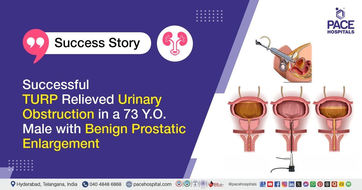
PACE Hospitals’ expert Urology team successfully performed a Transurethral Resection of the Prostate (TURP) on a 73-year-old male patient diagnosed with benign prostatic enlargement, lower urinary tract symptoms, and acute urinary retention requiring urinary catheterisation. The procedure aimed to remove the obstructing prostate tissue, restore normal urine flow, relieve urinary symptoms, and reduce the risk of bladder and kidney-related complications.
Chief Complaints
A 73-year-old male patient with a body mass index (BMI) of 20 presented to the Urology Department at PACE Hospitals, Hitech City, Hyderabad, with complaints of reduced urine flow for the past three months. His urinary symptoms had progressively worsened, and he developed acute urinary retention requiring urinary catheterization.
Past Medical History
The patient had no documented significant medical comorbidities, previous urological procedures, major surgeries, or chronic illnesses. The patient had no reported history of diabetes mellitus, hypertension, chronic kidney disease, cardiac disease, respiratory illness, neurological disorders, or known medication allergies.
On Examination
On examination, the patient was conscious, coherent, oriented, and hemodynamically stable. General physical examination showed no pallor, jaundice, cyanosis, clubbing, lymph node enlargement, or swelling.
Abdominal examination revealed a soft and non-distended abdomen without significant tenderness. There were no clinical signs of active urinary tract infection or dehydration. Cardiovascular, respiratory, and neurological examinations were within normal limits.
Diagnosis
Upon admission to PACE Hospitals, following a detailed clinical assessment, the Urology team evaluated the patient for reduced urine flow, lower urinary tract symptoms, and acute urinary retention requiring urinary catheterisation.
Ultrasound and transrectal ultrasound examinations were performed to assess the prostate gland, bladder outlet, and urinary tract. Based on the patient’s symptoms, clinical findings, and diagnostic investigations, he was diagnosed with benign prostatic enlargement associated with lower urinary tract symptoms and acute urinary retention.
Benign prostatic enlargement, also known as benign prostatic hyperplasia, is a non-cancerous enlargement of the prostate gland. As the prostate increases in size, it may compress the urethra and obstruct the normal passage of urine. This can cause reduced urine flow, difficulty starting urination, straining, incomplete bladder emptying, increased urinary frequency, urgency, night-time urination, and urinary retention.
Routine investigations, including complete blood picture, serum electrolytes, kidney and liver function tests, urine examination, urine culture, coagulation profile, blood grouping, blood sugar testing, viral screening, and pre-anaesthetic assessment, were performed to evaluate the patient’s overall fitness for surgery. The findings were within acceptable limits and supported proceeding with surgical treatment.
Based on the confirmed diagnosis, the patient was advised to undergo Benign Prostatic Enlargement Treatment in Hyderabad, India, under the expert care of the Urology Department.
Medical Decision Making (MDM)
After a detailed consultation with Consultant Laparoscopic Urologists Dr. Abhik Debnath, Dr. K. Ravichandra, and Senior Consultant Urologist Dr. Vishwambhar Nath, a comprehensive evaluation was performed to determine the most appropriate treatment approach.
Considering the patient’s reduced urine flow, lower urinary tract symptoms, acute urinary retention requiring catheterisation, enlarged 60-gram prostate, and prominent median lobe causing bladder outlet obstruction, the team assessed the severity of the urinary blockage and the possibility of continued catheter dependence.
Routine investigations, including complete blood picture, kidney function tests, urine examination, urine culture and sensitivity, blood sugar, liver function tests, serum electrolytes, and blood clotting tests, showed acceptable parameters for surgery.
Based on the patient’s symptoms, prostate size, imaging findings, catheter-dependent urinary retention, and preoperative investigations, it was determined that Transurethral Resection of the Prostate under spinal anaesthesia was the most appropriate and effective management strategy.
The procedure was selected to remove the obstructing prostate tissue, widen the urinary passage, improve urine flow, help the patient pass urine without a catheter, and reduce the risk of recurrent urinary retention and other urinary complications.
The patient and his family members were counselled regarding the diagnosis, investigation findings, planned surgical procedure, type of anaesthesia, expected recovery, possible risks, catheter care, postoperative urinary symptoms, and the importance of regular follow-up.
Surgical Procedure
Following the decision, the patient was scheduled for Transurethral Resection of the Prostate (TURP) in Hyderabad at PACE Hospitals under the expert care of the Urology Department.
The following steps were carried out during the procedure:
- Patient Preparation and Anaesthesia: After obtaining informed consent and completing the pre-anaesthetic evaluation, the patient was taken to the operating room. Under strict aseptic precautions, spinal anaesthesia was administered, and the patient was positioned appropriately.
- Insertion of the Resectoscope: A specialised endoscopic instrument called a resectoscope was gently passed through the urinary opening and advanced through the urethra towards the prostate. No external surgical incision was required.
- Identification of the Obstructing Prostate Tissue: The enlarged prostate tissue, including the prominent median lobe causing obstruction at the bladder outlet, was carefully identified.
- Resection of the Prostate Tissue: The obstructing portion of the prostate was gradually removed using an electrical cutting loop. The resected prostate tissue was flushed into the bladder and collected for histopathological examination.
- Control of Bleeding: Bleeding points within the operated area were carefully controlled to reduce the risk of postoperative bleeding.
- Catheter Placement and Completion: A Foley urinary catheter was inserted into the bladder to allow continuous drainage of urine and support healing of the operated area. The procedure was completed successfully without any intraoperative complications.
Postoperative Care
The patient had an uneventful postoperative recovery and remained hemodynamically stable throughout the hospital stay. He was regularly monitored for urine output, bleeding, hydration, kidney function, pain, and overall recovery. Appropriate treatment was provided to prevent infection, relieve pain and discomfort, protect the stomach, prevent constipation, and support recovery. The urinary catheter was removed on the third postoperative day, after which the patient passed urine comfortably with an improved urinary stream. He was gradually started on oral fluids and a normal diet and was discharged in a stable condition with postoperative instructions.
Discharge Medications
Upon discharge, the patient was prescribed oral medications to help prevent postoperative infection, reduce pain and discomfort, control fever when required, protect the stomach from irritation, prevent constipation, and support recovery following surgery. The patient was also advised to continue his routine medications as recommended.
Advice on Discharge
The patient was advised to drink adequate fluids, follow a balanced fibre-rich diet, avoid constipation and straining, and perform Kegel exercises as instructed. He was also advised to avoid heavy lifting, strenuous exercise, cycling, prolonged travel, and activities that place pressure on the pelvic region during the initial recovery period.
Emergency Care
The patient was advised to contact the emergency ward at PACE Hospitals in case of any emergency or development of symptoms such as fever, severe pain, vomiting, difficulty passing urine, reduced urine output, heavy bleeding, blood clots, dizziness, or worsening urinary symptoms.
Review and Follow-up Notes
The patient was advised to return for a follow-up visit with the Urologist in Hyderabad at PACE Hospitals after one week.
Conclusion
This case highlights the successful use of TURP to relieve urinary obstruction caused by benign prostatic enlargement. The procedure improved urine flow, allowed successful catheter removal, and supported comfortable urination. Appropriate postoperative care, Kegel exercises, and regular follow-up help promote recovery and improve urinary control.
Importance of Kegel Exercises After Transurethral Resection of the Prostate
Kegel exercises help strengthen the pelvic floor muscles that support the bladder and control urination. After TURP, some patients may temporarily experience urinary urgency, frequent urination, dribbling, or mild urine leakage. Regular pelvic floor exercises may improve bladder control, reduce leakage, and support the return of normal urinary function. These exercises involve tightening and relaxing the muscles used to stop urine flow. They should be started only when advised by a
urologist/urology doctor, usually after removal of the urinary catheter. Correct techniques and regular practice can support recovery and improve urinary control after TURP.
Frequently Asked Questions (FAQs)
Why was TURP advised for this patient?
TURP was advised because the patient had an enlarged prostate causing significant obstruction to urine flow. He had reduced urine flow, lower urinary tract symptoms, and acute urinary retention requiring catheterisation. Imaging also showed a 60-gram prostate with a large median lobe obstructing the bladder outlet. TURP was performed to remove the obstructing tissue and help him pass urine normally without a catheter.
What are lower urinary tract symptoms?
Lower urinary tract symptoms include problems related to storing or passing urine. They may include reduced urine flow, difficulty starting urination, straining, interrupted urine flow, incomplete bladder emptying, frequent urination, urgency, and repeated urination at night. These symptoms may occur due to prostate enlargement, bladder dysfunction, urinary infection, or other urinary conditions.
What happens during a TURP procedure?
During TURP, the urologist passes a specialised endoscopic instrument through the urinary opening into the urethra. The portion of the prostate blocking urine flow is gradually removed using an electrical cutting loop. The removed tissue is collected and sent for laboratory examination. A urinary catheter is placed temporarily to drain urine and allow the operated area to heal.
Is TURP an open surgery?
No. TURP is performed through the natural urinary passage and does not require an external abdominal incision. A specialised instrument is passed through the urethra to reach and remove the obstructing prostate tissue. This generally allows a shorter hospital stay and faster recovery than traditional open prostate surgery.
Why was a urinary catheter kept after TURP?
A urinary catheter helps drain urine while the operated prostate area heals. It also allows blood and small tissue particles to pass out of the bladder and helps prevent urinary blockage. Bladder irrigation may be provided through the catheter if required. The catheter is removed once the urine becomes clearer and the patient is considered ready to pass urine normally.
Is blood in the urine normal after TURP?
A small amount of blood in the urine may occur during the early recovery period after TURP. The urine may appear pink or light red, particularly after increased physical activity. This usually improves with rest and adequate fluid intake. However, heavy bleeding, large blood clots, dizziness, or inability to pass urine requires immediate medical attention.
Why is the removed prostate tissue sent for biopsy?
The prostate tissue removed during TURP is examined under a microscope through histopathological testing. This confirms the nature of the prostate enlargement and checks for any unexpected abnormal or cancerous cells. Although TURP is usually performed for benign prostate enlargement, laboratory examination of the removed tissue is an important part of postoperative evaluation.
How do Kegel exercises help after TURP?
Kegel exercises strengthen the pelvic floor muscles that support bladder control. They may help reduce temporary urine leakage, urgency, and dribbling after catheter removal. The exercises involve tightening and relaxing the muscles used to control urine flow. They should be performed regularly using the technique advised by the healthcare team.
When can normal activities be resumed after TURP?
Light walking and routine household activities can usually be resumed gradually as the patient feels comfortable. Heavy lifting, strenuous exercise, cycling, and activities that place pressure on the pelvic area should be avoided during the early recovery period. The urologist will advise when the patient can safely return to work, driving, exercise, and other normal activities.
Why should constipation and straining be avoided after TURP?
Straining during bowel movements increases pressure in the pelvic region and may cause bleeding from the healing prostate tissue. Constipation can also increase pain and discomfort during recovery. Drinking adequate fluids, eating fibre-rich foods, remaining gently active, and using prescribed bowel-supportive treatment can help prevent constipation.
Share on
Request an appointment
Fill in the appointment form or call us instantly to book a confirmed appointment with our super specialist at 04048486868
Appointment request - health articles
Recent Articles