Urethral Diverticulum - Symptoms, Causes, Prevention, Treatment
PACE Hospitals
Urethral diverticulum is a medical disorder that affects the urethra - the tube through which urine exits the body. This condition refers to an unusual pouch or sac-like structure that develops in the urethra. This disorder develops when there is a weakening or defect in the urethral wall, causing it to expand outward and produce a pocket. A urethral diverticulum can be congenital (existing at birth) or acquired later in life because of trauma or infections.
Urinary frequency, urgency, and pain while urinating are all common signs of urethral diverticula. Some people may also have recurring urinary tract infections or difficulty emptying their bladder. Urethral diverticulum is often diagnosed using a combination of past medical history, physical examination, and imaging techniques such as ultrasound and magnetic resonance imaging (MRI). The patient's treatment options differ depending on the degree and symptoms they experience. In rare circumstances, antibiotic treatment alone may be sufficient.
Proper management of the urethral diverticulum, including timely medical attention and potential surgical intervention by an expert urologist, can significantly improve an individual's quality of life. This emphasis on the potential for improvement can help people feel hopeful and optimistic about their condition.
Urethral diverticulum definition
Urethral diverticulum is defined as a medical condition characterized by the formation of a pouch or sac-like outpouching in the urethra. Urine can accumulate in the pouch and can cause infections.
Urethral diverticulum meaning
In 1805, William Hay- an English surgeon first documented a female with a sub-urethral diverticulum in the medical literature.
Dorland's Medical Dictionary defines a
diverticulum
as "a pouch or sac occurring normally or created by herniation (protrusion of an organ) of the lining mucous membrane through a defect in the muscular coat of a tubular organ."
Prevalence of urethral diverticulum
Urethral diverticula are estimated to affect 1-6% of women, with stress incontinence being the most common cause. Women experience urinary diverticula significantly more commonly than men do.
Although it can affect people of all ages, patients often present between the third and fifth decades of life. An incidence of 17.9 per 10,00,000 is reported annually for urethral diverticulum (UD). When women with
urinary incontinence (loss of bladder control) visit urology practices, urethral diverticulum is estimated to account for 1.4% of cases. The average age of diagnosis of urethral diverticulum is 45 years. This condition is rare in children.
Classification of urethral diverticulum
Leach- a urologist, and colleagues have proposed a classification system for urethral diverticula, which has not been generally used. The L/N/S/C3 classification system is a staging system for urethral diverticula that is based on various characteristics of the condition, such as location, number, size, anatomic configuration, site of communication to the urethral lumen, and patient continence status. It is comparable to the staging system used for cancer staging. Although other researchers have not applied or validated this system, it aims to standardize the description of urethral diverticula.
Another proposed classification scheme makes the location of the urethral diverticula the key determinant of surgical technique, with distal lesions undergoing marsupialization (Surgical treatment that creates a permanent hole in a cyst or abscess to allow fluid to drain) and more proximal lesions receiving excision and repair.
Another classification system for urethral diverticula by Leng and McGuire splits them into two groups based on whether or not the periurethral fascial layer remains intact. Some patients with urethral diverticula who have had past vaginal or urethral surgery may have a defective periurethral fascial layer, resulting in a pseudodiverticulum. These researchers argue that recognizing this anatomic arrangement has significant consequences for surgical reconstruction. Patients may require additional reconstruction or the placement of a tissue flap or graft to strengthen the repair and prevent recurrence or postoperative urethrovaginal fistula formation.
Urethral diverticulum symptoms
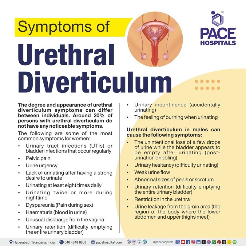
The degree and appearance of urethral diverticulum symptoms can differ between individuals. Around 20% of people with urethral diverticulum do not have any noticeable symptoms. The following are some of the most common symptoms for women:
- Urinary track infections (UTIs) or bladder infections that occur regularly
- Pelvic pain
- Urine urgency
- Inability to urinate after having a strong desire of urination
- Urinating at least eight times daily
- Urinating twice or more during the night
- Dyspareunia (Pain during sex)
- Haematuria (blood in urine)
- Unusual discharge from the vagina
- Urinary retention (difficulty emptying the entire urinary bladder)
- Urinary incontinence (accidentally urinating)
- Feeling of burning when urinating
Urethral diverticulum in males can cause the following symptoms:
The unintentional loss of a few drops of urine while the bladder appears to be empty after urinating (post-urination dribbling)
- Urinary hesitancy (difficulty urinating)
- Weak urine flow
- Abnormal size of penis or scrotum
- Urinary retention (difficulty emptying the entire urinary bladder)
- Restriction in the urethra
- Urine leakage from the groin area (the region of the body where the lower abdomen and upper thighs meet)
Urethral diverticulum causes
Causes of congenital diverticulum
It is demonstrated that urethral diverticula are either congenital (a condition or trait that exists by birth) or acquired (developed or originating after birth).
Although congenital urethral diverticula are uncommon, the congenital origin argument has been supported by cases of urethral diverticula in children. The following conditions are said to be cause of congenital urethral diverticula:
- Remnant of Gartner duct
- Faulty union of primordial folds
- Vaginal wall cysts of Mullerian origin
- Congenital dilatation of periurethral cysts
- Association with blind-ending ureters
Remnant of Gartner duct: Gartner's duct remnants are vestigial remnants of the mesonephric ducts that typically regress after the eighth week of embryonic development.
Faulty union of primordial folds: “Faulty union of primordial folds” refers to a congenital condition in which embryonic structures fuse or join abnormally during development. Especially in the context of urethral diverticula.
Vaginal wall cysts of Mullerian origin: Vaginal wall cysts of Müllerian origin are a common type of vaginal cyst that arises from tissues left behind during pregnancy. They can grow anywhere on the vaginal wall and frequently contain mucous. They are often small and asymptomatic, but they can grow large enough to induce pressure or heaviness on adjacent structures.
Congenital dilatation of periurethral cysts: Congenital dilatation of periurethral cysts, often known as congenital paraurethral cysts (CPUCs), is a rare disease that affects women. It is caused by a blockage in the ducts of the paraurethral glands, which function similarly to the male prostate.
Association with blind-ending ureters: A blind-ending ureter is an uncommon congenital urinary abnormality known as a ureteral duplication anomaly (defect). It is caused by premature division of the ureteric bud during development.
Causes of acquired urethral diverticulum
Acquired urethral diverticula are brought on by recurrent urinary tract infections that block the periurethral glands (tiny glands that open into the female urethra) and can burst into the urethral lumen, resulting in local herniation (protrude through an abnormal body opening). The role of the periurethral glands in the development of urethral diverticulum is significant. When these glands become obstructed due to infections, they can fill with fluid or other substances and eventually protrude into the urethra, leading to the formation of a diverticulum.
Urethral diverticulum risk factors
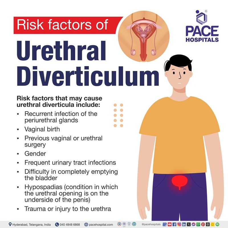
Risk factors that may cause urethral diverticula include:
- Recurrent infection of the periurethral glands
- Vaginal birth
- Gender
- Frequent urinary tract infections
- Difficulty in completely emptying the bladder
- Hypospadias (condition in which the urethral opening is on the underside of the penis)
- Trauma or injury to the urethra
Recurrent infection of the periurethral glands
The majority of urethral diverticula are thought to be caused by periurethral gland infections that occur repeatedly or over time. The sub-urethral infection can clog the ducts and glands, resulting in cystic expansion and retention cysts (cysts that forms due to blocked glands).
Vaginal birth
One possible risk factor for urethral diverticulum development is vaginal delivery of the baby. This is because the urethral muscles and glands are under stress during childbirth.
Gender
As women are more likely to develop urethral diverticulum, gender might increase the chances of developing urethral diverticulum.
Frequent urinary tract infections (UTIs)
This condition is likely caused by recurring urinary tract infections (UTIs) that block the periurethral glands surrounding the urethra. Urinary tract infections can cause the glands to become obstructed, fill with fluid or other substances, and eventually protrude into the urethra.
Difficulty in completely emptying the bladder
Urinary tract infections may occur as a complication in people suffering from urinary retention (the inability to empty the bladder). Recurrent infections may lead to weakness in the urethral wall, eventually forming a urethral diverticulum.
Hypospadias (a condition in which the urethral opening is on the underside of the penis)
It is demonstrated that urethral diverticula may be formed as a result of hypospadias complications.
According to research, this complication accounts for 0.3% of postoperative complications and is most commonly related to proximal hypospadias.
Trauma or injury to the urethra
Trauma can cause urethral diverticulum (UD). There have been cases reported on urethral diverticulum due to urethral trauma.
Complications of urethral diverticulum
Urethral diverticulum is a rare disorder that can cause a variety of consequences. Complications linked to urethral diverticulum may include:
- Abscess (buildup of pus)
- Urethral calculi (stones)
- Neoplasm (cancer)
- Recurrent diverticulum (the return of urethral diverticulum after a period of remission)
- Stress incontinence (bladder leakage due to physical activity or exertion)
- Urethral stricture (scarring of tissue in the urethra)
- Recurrent urinary tract infections

Prevention of urethral diverticulum
Urethral diverticula (UD) are not always prevented. However, specific steps may help to reduce the chances of the development of urethral diverticulum, including:
- Appropriate treatment of urinary tract infections: Recurrent or chronic urinary tract infections (UTIs) can cause inflammation and weakening of the urethral tissues, increasing the risk of diverticulum formation. Hence, managing urinary tract infections promptly may help prevent urethral diverticulum.
- Involving in pelvic floor exercises: Pelvic floor exercises, often known as Kegel exercises, can help strengthen the muscles that support the urological system and enhance urologic health.
- Avoidance of trauma: Urological trauma, also known as urotrauma, is a traumatic incident or impact that causes damage to the urinary tract or reproductive organs. It can be caused by a fall, a vehicle or bicycle accident, a chemical, or a weapon. Injuries to the urinary tract may create complications such as urethral diverticulum.
- Ensure good hygiene: Drinking a lot of water, maintaining a healthy weight, and avoiding acidic foods, excessive alcohol and caffeine, and other foods and beverages that can irritate the urethra may help keep the urinary system healthy.
Diagnosis of urethral diverticulum
Although urethral diverticulum is usually difficult to diagnose, it has been detected more frequently in recent decades as physicians have become more aware of the condition. Here's an overview of the steps involved in diagnosing urethral diverticulum:
- Medical history
- Physical examination
- Imaging Studies (urethral diverticulum radiology): The primary imaging investigations utilized in the diagnosis of urethral diverticula include:
- Voiding cystourethrography
- Magnetic resonance imaging (MRI)
- Ultrasonography
- Intravenous pyelography and computed tomography (CT) urography
- Retrograde urethrography using a double-balloon catheter
- Urine culture
- Procedures:
- Urodynamic studies
- Cystoscopy
Urethral diverticulum treatment
Currently, there is no known medical therapy for successfully treating urethral diverticulum. Long-term low-dose antibiotic therapy may alleviate localized symptoms, but the structural abnormalities persist. Surgery is the current therapy of choice. Patients with substantial symptoms such as recurring urinary tract infections, severe discomfort, dyspareunia, frequency, urgency, and postvoid dribbling may correct their urethral diverticula surgically. Surgery is also indicated for urethral calculi, urinary retention, and cancer.
Before surgery, infected urethral diverticula are treated with suitable antibiotics. Surgical intervention is not possible until an active urinary tract or diverticulum infection is eliminated.
In the case of a urethral abscess, it may be treated by transvaginal aspiration or incision and drainage under examination or with ultrasonographic guidance before the actual therapy.
Surgical interventions
Several open surgical and endoscopic techniques have been described for treating urethral diverticula, including the following:
- Transurethral cauterization of the diverticulum
- Marsupialization of the diverticular sac into the vagina
- Excision of the diverticulum
Difference between cystourethrocele and urethral diverticulum
Cystourethrocele vs Urethral diverticulum
Understanding the variations between cystourethrocele and urethral diverticulum is critical for a correct diagnosis and appropriate therapy. Both disorders predominantly affect the female urinary system, although they have different characteristics, causes, and symptoms. Exploring these variations allows the urologists to better understand the unique challenges that each condition offers, as well as the specialised management approaches that are required.
| Aspect | Cystourethrocele | Urethral Diverticulum |
|---|---|---|
| Meaning | Protrusion of the bladder and urethra into the vagina | Outpouching or sac-like structure in the urethral wall |
| Location | Typically, the anterior vaginal wall near the urethra | Adjacent to the urethra |
| Causes | Weakness of pelvic floor muscles | Structural defect or obstruction |
| Symptoms | Pelvic pressure, Urinary incontinence and Difficulty urinating | Dysuria, Frequency and urgency and Recurrent urinary tract infections |
| Diagnosis | Pelvic examination, imaging (ultrasound, MRI) | Imaging (MRI, cystoscopy) |
| Treatment | Pelvic floor exercises, pessaries, surgery | Antibiotics for infections and surgical excision |
Frequently Asked Questions (FAQs) on Urethral diverticulum
Is the urethral diverticulum cancerous?
Yes, urethral diverticula (UD) are linked to urethral cancer, with a 1-8% probability of developing the disease. Urethral diverticulum carcinoma (UDC) is an uncommon disorder, accounting for about 30% of urethral malignancies that develop from a urethral diverticulum.
Is the urethral diverticulum serious?
Yes. The urethral diverticulum can be severe if not treated or managed properly, problems such as recurring infections that cause kidney damage, urine retention (inability to fully empty the bladder), fistula formation (abnormal connections between organs), and persistent pelvic pain can develop.
Is surgery necessary for the urethral diverticulum?
Yes. In cases of a painful urethral diverticulum, Surgery is necessary to treat urethral diverticulum (UD) effectively. In other circumstances, non-surgical therapy may be sufficient, such as monitoring, antibiotics, or urethral dilation and massage.
At what age urethral diverticulum is commonly diagnosed?
It is rare but more common in women aged 40 to 70. Children are normally unaffected unless they have undergone urethral surgery. With improved imaging, more urethral diverticula have been discovered and treated.
Can the urethral diverticulum reoccur after surgery?
Female urethral diverticulum is a rare disorder that affects 1–6% of women. Recurrence following surgery is standard, with a prevalence ranging from 8 to 20%. It typically affects women between their third and seventh decades of life, but it can occur at any age. Typically, it appears as a protrusion beneath the urethra lined with urethral mucosa.
What is urethral diverticulum?
A urethral diverticulum is a disorder in which a fluid-filled sac develops on the urethra, which drains urine from the body. These sacs can get contaminated, resulting in various disorders.
What is anterior urethral diverticulum?
Anterior urethral diverticula develop when a pocket or sac forms along the anterior (front) wall of the urethra. The diverticulum can be found anywhere throughout the anterior urethra; however, it is most typically observed in the distal bulbar and proximal penile urethra.
What is the posterior urethral diverticulum?
Urethral diverticula are known as posterior diverticula because they typically originate on the posterior aspect of the urethra, which is the tube that carries urine out of the body. They are saccular urethral mucosal evaginations (turning outward or inside out) that protrude posteriorly into the urethrovaginal region.
What is the gold standard urethral diverticulum diagnostic test?
Magnetic resonance imaging (MRI) is the gold standard for diagnosing urethral diverticula (UD) because it produces crisp images of the body without the use of X-rays. MRI can detect the size and location of the diverticulum and is more sensitive than other imaging procedures such as voiding cystourethrogram (VCUG). MRI can be utilised for staging and surveillance.
How is urethral diverticulum formed?
A urethral diverticulum develops when urine accumulates in the sub urethral cyst during urination on multiple occasions. Urethral diverticula are typically found in the distal part of the urethra. Although uncommon, distal urethral diverticula can also be caused by a blocked Skene gland.
Is the urethral diverticulum a cyst?
A urethral diverticulum manifests as a cystic lesion usually appearing in the posterolateral mid/distal urethra. It usually coils around the urethra. It could be multilocular (split into several tiny compartments or vesicles).
What can be mistaken for urethral diverticulum?
Symptoms of urethral diverticulum are frequently confused with disorders such as, endometriosis, gartner duct cyst, interstitial cystitis, mullerian duct anomalies, overactive bladder, pelvic inflammatory disease, skene gland abscess, ureterocele, urethral cancer, vaginal inclusion cyst.
What are the three D's of the urethral diverticulum?
The traditional presentation of UD is known as the "3 D's":
- Dysuria (painful urination)
- Dyspareunia (painful intercourse)
- Drip of urine following urination (involuntary loss of urine)
However, these three symptoms are only sometimes present in many patients with urethral diverticulum and co-occur in only around 20% of all instances.
Related articles
Share on
Request an appointment
Fill in the appointment form or call us instantly to book a confirmed appointment with our super specialist at 04048486868

