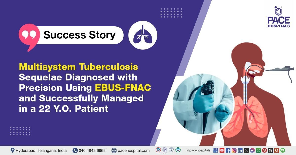
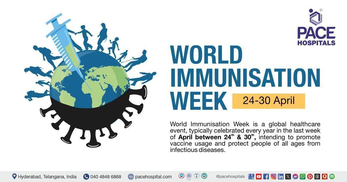
Successful Closed Reduction and K-Wire Fixation of a Pediatric Supracondylar Humerus Fracture
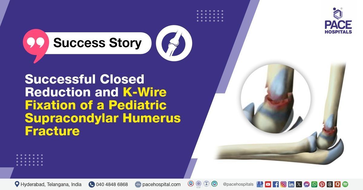
The orthopedic team at PACE Hospitals successfully performed a closed reduction and K-wire fixation procedure for a left supracondylar humerus fracture involving the elbow in a 5-years-old female patient with complaints of pain, deformity, and swelling in the left elbow following a fall on the playground.
Chief Complaints
A 5-year-old female child was brought to
PACE Hospitals, Hitech City, Hyderabad, by her parents with complaints of pain, visible deformity, and swelling in the left elbow following a fall while playing on the playground. The injury occurred due to a sudden tumble during play, causing significant discomfort and distress.
History of present illness
The patient presented with pain, deformity, and swelling in the left elbow following a fall while playing on the playground. Due to the severity of the pain, the patient also reported difficulty in using the left upper limb for daily activities.
General examination
Upon admission to PACE Hospitals, the patient's vital signs were stable. On general examination, the patient appeared alert and oriented. Local examination of the left elbow revealed swelling, tenderness, and visible deformity. Range of motion was painfully restricted, but there were no signs of distal neurovascular deficit.
Diagnosis
After being admitted to PACE Hospitals, the orthopaedic team thoroughly reviewed the patient's medical history and conducted a detailed clinical examination. Based on the presenting symptoms and physical findings, there was a strong clinical suspicion of a left supracondylar humerus fracture.
The diagnosis of a supracondylar humerus fracture was confirmed through clinical evaluation and imaging prior to the intervention.
Based on the diagnosis and the complex presentation of the condition, she was advised to undergo for a Left Supracondylar Humerus Fracture Treatment in Hyderabad, India, under the care of the Orthopaedic Department, ensuring comprehensive management of the fracture.
Medical Decision-Making
Considering the patient's pain, difficulty in carrying out daily tasks, and the diagnosis, it was determined that surgical intervention was necessary. After a thorough discussion with the patient's guardians and obtaining their informed consent, Dr. Raghuram, Consultant Orthopaedic Surgeon, decided to proceed with a Closed Reduction and K-Wire Fixation procedure for the left supracondylar humerus fracture involving the elbow.
Surgical Procedure
Following the decision, the patient was scheduled for a Closed Reduction and K-Wire Fixation procedure in Hyderabad at PACE Hospitals, under the expert supervision of the Orthopaedic Department, for the management of a left supracondylar humerus fracture involving the elbow.
The patient underwent closed reduction and K-wire fixation under general anaesthesia. During her hospital stay, she remained stable and was closely monitored.
Treatment Considerations
Closed Reduction and K-Wire Fixation is commonly considered in paediatric patients to realign fractured bones without the need for large incisions. This is followed by the insertion of Kirschner wires (K-wires) to stabilize the bone and maintain proper alignment throughout the healing process.
This technique is particularly effective for supracondylar humerus fractures, which are frequently seen in children. It offers several advantages, including minimal soft tissue disruption, reduced risk of complications, and accelerated healing. Being a minimally invasive method, it also helps preserve the growth plate. The procedure is especially beneficial for displaced fractures that are not extensively comminuted, enabling quicker recovery and easier rehabilitation.
However, the decision to proceed with this approach depends on various factors such as the type and severity of the fracture, the patient’s age, and potential risks related to healing. Post-operative monitoring remains essential to detect and manage any complications, particularly neurovascular compromise or restricted elbow mobility.
Postoperative Care
The patient’s immediate postoperative recovery was uneventful, with no major complications. Postoperative care included antibiotics, analgesics, and supportive measures.
Because the fracture was comminuted, the risks of malunion and deformity were thoroughly explained to the patient and her family. Counselling was provided on the importance of follow-up visits and rehabilitation.
Discharge notes
The patient was discharged in a stable condition.
Discharge Medications
Upon discharge, the patient was prescribed a non-steroidal anti-inflammatory drug (NSAID) to alleviate pain and reduce inflammation, along with dietary supplements to promote bone healing and support nutritional recovery.
These medications were prescribed to ensure comfort during rehabilitation and provide the nutrients needed for optimal fracture repair.
Advice on Discharge
The parents were instructed to perform active finger exercises to preserve circulation and prevent joint stiffness. An arm sling was provided for support, and limb elevation was recommended to minimise swelling.
Emergency Care
The parents was informed to contact the Emergency ward at PACE Hospitals in case of any emergency or development of symptoms like fever, abdominal pain, or vomiting.
Review and follow-up notes
The parents were advised to return to the Orthopedic Department at PACE Hospitals in Hyderabad for a follow-up appointment after one week. During this visit, a comprehensive evaluation, wound assessment, and monitoring of postoperative recovery would be performed.
Conclusion
This case underscores the value of collaborative care in the evaluation and management of a left supracondylar humerus fracture (which involved the bone breaking into multiple fragments) in a child.
Importance of Proper Management and Classification in Paediatric Supracondylar Humerus Fractures
Supracondylar humerus fractures are among the most common childhood injuries, particularly affecting children between the ages of 5 and 8 years. The most frequent type is the extension fracture, often with posteromedial displacement. While orthopedists/orthopedic surgeons agree on some fundamental principles, there is still ongoing debate about the best way to classify these fractures, when to realign the bone, and which cases require urgent intervention.
There are questions regarding the safest methods of fracture reduction, with disagreement on whether closed or open reduction is more effective, as well as the optimal placement of stabilising pins. The use of a medial pin, in particular, remains a point of contention. Moreover, managing cases where the hand appears pink but lacks a pulse is critical, as well as understanding how much the growing bone can naturally remodel and the long-term risks of cubitus varus, where the elbow heals slightly crooked. Gartland’s classification provides valuable guidance in determining treatment strategies, with closed reduction and percutaneous pinning being the preferred approach. Open reduction is typically reserved for more complex or failed cases. When managed appropriately, even fractures involving vascular or nerve injuries can result in favourable outcomes, demonstrating the importance of timely and effective treatment.
Share on
Request an appointment
Fill in the appointment form or call us instantly to book a confirmed appointment with our super specialist at 04048486868
Appointment request - health articles