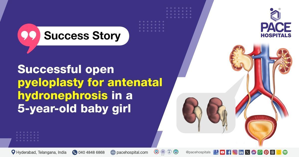
Successful open pyeloplasty for antenatal hydronephrosis in a 5-year-old baby girl | Case study
PACE Hospitals
PACE Hospitals' Urology team successfully performed a right open pyeloplasty (dismembered) for a 5-year-old baby girl with right pelviureteric junction (PUJ) obstruction and with a history of right hydronephrosis before birth (Antenatal hydronephrosis).
A 5-year-old baby girl with complaints of on and off-right flank pain presented to the consultant laparoscopic urologist, Dr. Abhik Debnath, at PACE Hospitals, Hitech City, Hyderabad.
Medical History
Delving deeper, it was understood that the patient was suffering from right flank pain (on and off) which led to her admission to the PACE Hospitals for additional care and management. The patient had a history of right hydronephrosis before birth (Antenatal hydronephrosis).
Diagnosis
Upon being admitted to PACE Hospitals and understanding the medical history, the patient was subjected to computed tomography urography, complete blood picture (CBP), kidney function tests including blood urea nitrogen (BUN), creatinine, glomerular filtration rate (GFR), urine culture and sensitivity, X-ray chest, basic metabolic panel (To assess the concentration of glucose, calcium, sodium, potassium, carbon dioxide, chloride). Based on the diagnostic investigations, the patient was diagnosed with:
Right pelviureteric junction (PUJ) obstruction with a history of right hydronephrosis before birth (ANH).
The PUJ is the junction between the pelvis of the kidneys and the ureter (the tube connecting the kidney to the bladder). Pelviureteric junction obstruction (PUJO) is a widely recognized clinical condition that causes impaired urine flow from the renal pelvis into the ureter and, if not observed and treated effectively, can result in a loss of the affected kidney. PUJO is generally a congenital condition and is the most prevalent cause of hydronephrosis identified before birth. It can be identified using antenatal ultrasound during the second trimester of pregnant women.
Antenatal hydronephrosis (ANH) is the most common congenital condition defined as enlargement of the renal pelvis and/or calyces in newborns. It is commonly detected during pregnancy using an antenatal ultrasound examination.
Treatment
After consultation with the team of consultant laparoscopic urologists that includes Dr. Abhik Debnath, and other senior consultant Urologists & Renal Transplant Surgeons, it was determined that the right open pyeloplasty (dismembered) surgery was the effective method for treating the patient. This minimally invasive open dismembered pyeloplasty is technically possible, and it is safe and successful treatment for ureteropelvic junction obstruction. It is performed quickly, with a small incision, fewer complications, and a short recovery period.
A day before open pyeloplasty (dismembered) surgery, urologists performed necessary diagnostic procedures, including computed tomography urography, complete blood picture (CBP), kidney function tests, urine culture and sensitivity, X-ray chest, basic metabolic panel tests to look for any abnormalities and to assess the severity of hydronephrosis caused by ureteropelvic junction (UPJ) obstruction.
Performing these procedures before open pyeloplasty (dismembered) surgery ensures that the urologists have a comprehensive understanding of the patient's condition and anatomy, which guides them in performing the surgery effectively and helps reduce the risk of complications.
With necessary investigations done & clearances obtained, which included a pre-anesthesia checkup, the patient underwent a right open pyeloplasty (dismembered) surgery under general anesthesia. The procedure was supervised by the consultant laparoscopic urologist – Dr. Abhik Debnath and it was accomplished devoid of any complications.
The aftermath
The post-operative period was uneventful. Regular tests were done to understand the overall health of the patient. Foley's catheter (a thin, flexible catheter to drain urine from the bladder) and the drainpipe were removed on postoperative day 3.
The necessary medicines, antibiotics, multivitamins, analgesics, proton pump inhibitors & other supportive care were given along with counselling. The patient was discharged upon achieving hemodynamic stabilization, with the necessary medications and was advised to continue routine medications, adequate fluids, and normal diet.
The patient was also instructed to contact PACE Hospitals in case of adverse symptoms like fever, abdominal pain, or vomiting. After 6-8 weeks, the patient was asked to get a review by Dr. Abhik Debnath for cystoscopy and ureteral stent (DJ Stent) removal.
Importance of radiologic imaging in diagnosing pelviureteric junction (PUJ) obstruction
Radiologic imaging plays an essential role in diagnosing PUJ obstruction. Congenital PUJ obstruction is the leading cause of upper urinary tract obstruction in children. Due to the increasing use of prenatal ultrasound imaging, ultrasonographic abnormalities are now the major presentation of congenital pelviureteric junction (PUJ) obstruction. Routine prenatal evaluations are normally performed between 16 and 20 weeks of gestation.
PUJ obstruction indicates functionally obstructed urine transport from the renal pelvis into the ureter as the increased renal pelvic pressure caused by blockage can lead to progressive renal damage. This, combined with the chronicity and severity of the problem, determines the course of treatment.
Angiography of the renal artery is often performed before surgery to define crossing or supernumerary arteries which may be causing extrinsic pelviureteric junction (PUJ) obstruction.
Share on
Request an appointment
Fill in the appointment form or call us instantly to book a confirmed appointment with our super specialist at 04048486868


