A challenging case of paediatric liver transplant and seizures | Case study
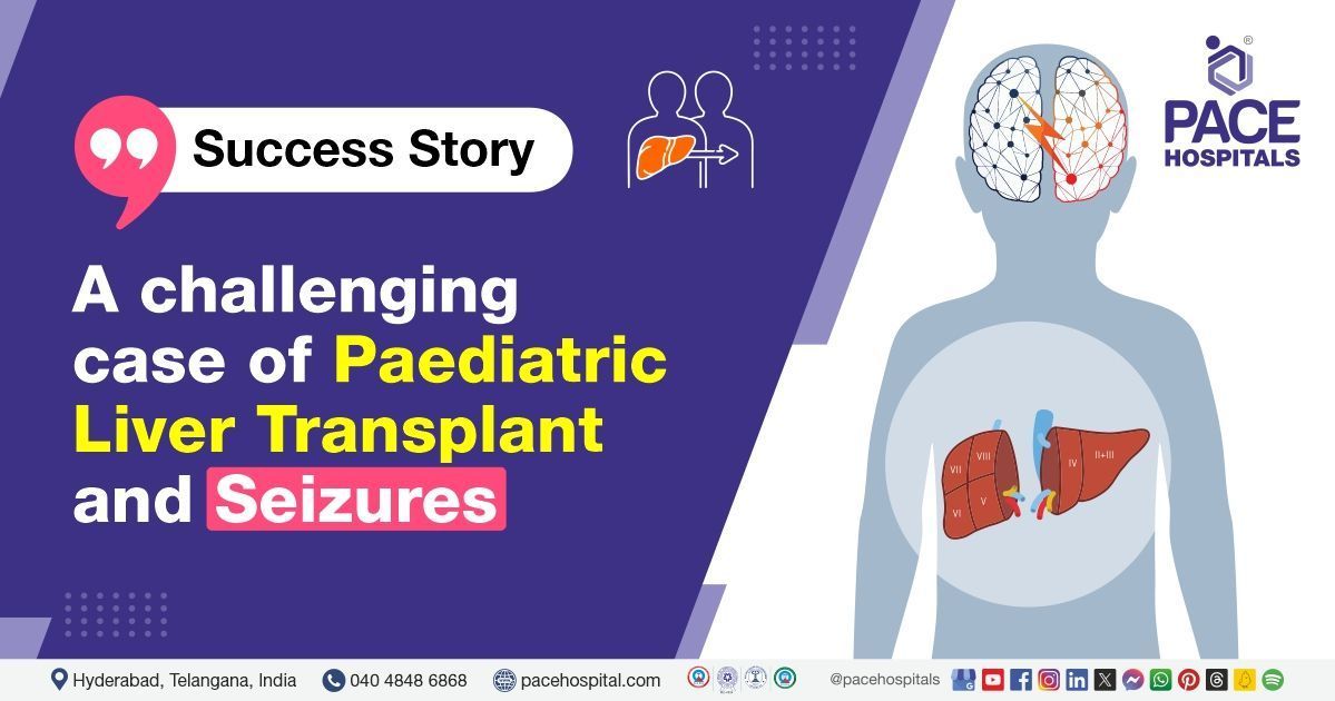
PACE Hospitals' Liver Transplant team successfully performed a Living Donor Liver Transplantation (LDLT) for a 9-month-old female infant with jaundice and intermittent fever for the past month.
A 9-month-old female infant (Priya) presented to the
PACE Hospitals, Hitech City, Hyderabad with complaints of jaundice for the past month, accompanied by intermittent fever.
Medical History
Delving deeper, it was understood that the patient had been experiencing jaundice for the past thirty days. The patient has a known history of extrahepatic biliary atresia type 1 (partial or total absence of permeable bile duct between the porta hepatis and the duodenum) as evidenced by the presence of jaundice, clay-coloured stool, and hepatomegaly (Enlarged liver) early in her life.
A little over eight months ago, this infant underwent Kasai’s procedure (also called Kasai hepatoportoenterostomy) and duodenojejunostomy to treat biliary atresia. At the time of Kasai’s procedure, it was reported that the patient was found with
- Lobular cholestasis (accumulation of bile within the hepatocytes (liver cells) in the lobules of the liver)
- Bile ductule proliferation (increased growth and multiplication of small tubes carrying bile - bile ductules commonly seen in biliary cirrhosis)
- Focal fibrosis (incomplete nodule formation)
Diagnosis
Upon being admitted to PACE Hospitals and after understanding the history and physical examination, the patient was subjected to various appropriate investigations, such as liver biopsy, among others. Evaluating the diagnostic results, the patient was diagnosed with:
Biliary atresia post Kasai status
Biliary atresia is a progressive cholangiopathy (chronic disease primarily affecting the cholangiocytes - the cells lining bile ducts) of early infancy, which could result in gradual scar formation and ultimately obliterating the bile ducts that results in biliary cirrhosis and death of the child, usually within the first two years of life.
The Kasai portoenterostomy (popularly called as/ the Kasai procedure) is a surgery to construct a proper drainage. Although a Kasai portoenterostomy (KPE) is a critical surgery that may accomplish the restoration of bile flow (if performed before three months of age), thus assisting in the prevention of rapid liver injury progression as well as cirrhosis, unfortunately, a majority of the affected paediatric cases eventually develop end-stage liver disease insidiously, reiterating the need for paediatric liver transplantation.
Based on the diagnosis, the patient was evaluated for progressive liver dysfunction and was advised to undergo Paediatric End-Stage Liver Disease Treatment in Hyderabad, India, under the specialized care of the Liver Transplant Department.
Treatment
The liver transplant doctors (Dr. CH. Madhusudhan, Dr. Govind Verma, and Dr. Suresh Kumar S) asserted that a liver transplant was the only way to salvage/save the patient.
The patient was kept on multiple lifesaving supports. Understanding the condition of the patient, her mother came forward wilfully to donate a portion of her liver.
With necessary investigations done & clearances obtained, which included a pre-anaesthesia checkup, the patient underwent
living donor liver transplantation (LDLT), receiving the left lateral segment. The procedure was supervised by the liver transplant surgeon, Dr Madhusudhan, and it was accomplished without any complications.
The aftermath
The postoperative period was uneventful, which can be evidenced by the results of liver Doppler test (a non-invasive test that studies the blood supply in and around the liver through ultrasound waves). Postoperatively, the liver transplant doctor/specialist shifted the patient to Surgical Intensive Care Unit (SICU) and was kept on mechanical ventilator.
Mechanical ventilation is the practice of assisted ventilation (breathing), especially in cases where the patients cannot respire on their own. A majority of paediatric liver transplant cases require immediate postoperative mechanical ventilation. There are various reasons for this practice which could include:
- Postoperative respiratory depression arising from opioids
- Any preexisting malnutrition
- Concerns about graft function
- Mismatch in the organ-recipient size
- Poor cooperation, especially in young children
By the 4th day after liver transplantation, the infant achieved relative stability, prompting extubation (removal of mechanical ventilator tube), and non-invasive ventilation was started. On the 6th day, the patient developed sudden seizures, presumably hypoxic seizures.
On an immediate basis, both a pediatrician and the neurologist were consulted, and, on their referral, the hypoxic seizures were managed with an anticonvulsant. Acknowledging the risk of hypoxia and the development of any such similar mishaps in the future, the infant patient was kept on a continuous high-flow nasal cannula (HFNC), a non-invasive ventilation practice which ensured the delivery of 100% humidified and heated oxygen at a flow rate of up to 60 liters per minute, thus avoiding hypoxic state. Upon stabilisation, the patient was shifted from the Surgical Intensive Care Unit (SICU) to the in-patient room on the 12th day.
On the 14th day, she developed continuous fever episodes; upon investigation, it was found that fluid had been collected in the right hypochondrium (RHC) (upper right abdominal area). In view of the suspected bile leak, the baby was reeled to Surgical Intensive Care Unit (SICU).
There, her therapeutic regimen was once again evaluated, resulting in the immediate upgradation of the antibiotics. Understandably, this upgradation of the antibiotics was done to reduce the chances of any infection in the bile leak and an ultrasound-guided percutaneous catheter drainage was inserted in the abdomen to manage it.
On the 16th day, a sudden respiratory distress development necessitated intubation and she underwent emergency bronchoscopy promptly. The bronchoscopy defined the presence of occasional mucoid secretions, and subsequently, they were cleared. Since she displayed a gradual symptomatic improvement in two days, she was extubated and again shifted to the in-patient room on the 19th day.
The necessary medicines, immunosuppressives, antibiotics, proton pump inhibitors, multivitamins, antiemetics, analgesics, antipyretics & other supportive care were given along with the counselling. Upon achieving hemodynamic stabilisation, the patient was discharged with the necessary medications. She was advised to schedule a follow-up appointment with the
Liver Transplant Surgeons in Hyderabad at PACE Hospitals to assess her post-operative condition and ensure continued recovery.
Conclusion
This case highlights the importance of early diagnosis, coordinated multidisciplinary care, and timely surgical intervention in achieving successful outcomes in complex cases involving seizures and liver failure. It underscores the success of Paediatric Liver Failure Treatment in Hyderabad, India, leading to significant recovery and improved quality of life for the patient.
The clinicopathologics of seizure development after liver transplant
Despite the development of significant strides and progress in the field of liver transplant, which exponentially increased its therapeutic effectiveness since its inception in 1963, research has not been completely able to negate all the complications arising from a liver transplant.
Neurological complications can affect up to one-third of liver-transplanted patients. Studies demonstrated a higher mortality rate, increased hospitalisation, and elevated frequency of infections along with a lower self-sufficiency and social reintegration among liver transplantation cases associated with the development of neurological complications when compared with liver transplantation cases not associated with neurological complications.
Seizures are one of the most common neurological complications reported among liver transplantation cases. Seizures can be partial or generalized, most frequently of the tonic-clonic type. They can occur with or without magnetic resonance imaging -detectable structural brain lesions and with or without electroencephalography alterations within the inter-critical period. Immediately after liver transplantation, their prevalence may reach 10–40%, later their prevalence decreases to about 10%.
Seizures are likely to be due to immunosuppressors' neurotoxicity, rapid electrolytes or osmolar changes, ischaemic brain lesions, and demyelination of any origin. Seizures can be isolated or be a sign of encephalopathy. Usually, epilepsy after liver transplantation is self-limiting, but some patients may also develop severe treatment-resistant epilepsy.
The incidence of seizures after liver transplantation has declined in recent years, possibly because of the remarkable improvement in the management of the multiple metabolic and toxic abnormalities that may predispose to seizures. In addition, most transplant centers now avoid intravenous cyclosporine loading, and both the use of tacrolimus and the monitoring of its plasma levels, thus contributing to the decreased rate of seizures.
Share on
Request an appointment
Fill in the appointment form or call us instantly to book a confirmed appointment with our super specialist at 04048486868

