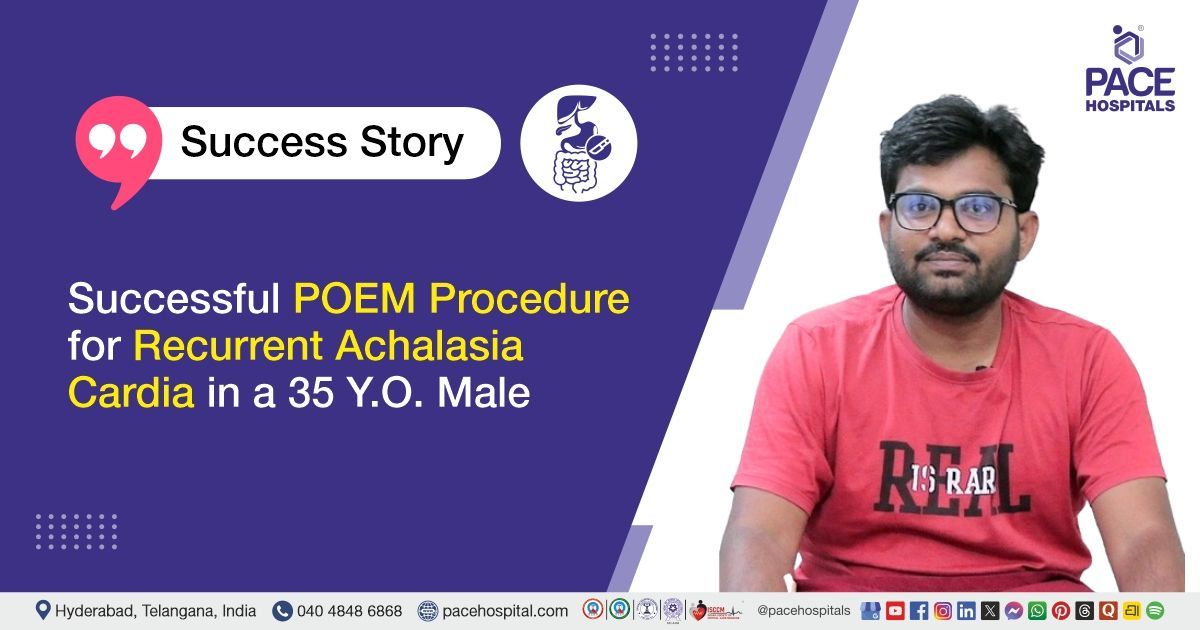
Successful Buccal Mucosal Graft Urethroplasty for Bulbar Urethral Stricture
PACE Hospitals
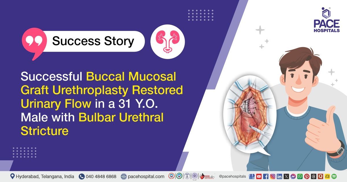
PACE Hospitals’ expert Urology team successfully performed Buccal Mucosal Graft Urethroplasty on a 31-year-old male patient diagnosed with a bulbar urethral stricture, chronic urinary retention, decompensated urinary bladder, and bilateral moderate hydroureteronephrosis. The procedure was performed to reconstruct the narrowed portion of the urethra, relieve the bladder outlet obstruction, improve urinary flow, and protect the upper urinary tract from further damage.
Chief Complaints
A 31-year-old male patient with a body mass index (BMI) of 22 presented to the Urology Department at PACE Hospitals, Hitech City, Hyderabad, with complaints of poor urinary flow and difficulty in passing urine.
Past Medical History
The patient had no known history of diabetes mellitus, hypertension, cardiac disease, chronic kidney disease, or any other significant medical condition. He had not undergone any previous major surgeries or urological procedures, and no other relevant past illnesses had been reported.
On Examination
On examination, the patient was conscious, coherent, oriented, and hemodynamically stable. Clinical assessment was suggestive of chronic bladder outlet obstruction with reduced urinary flow. Abdominal examination did not reveal any acute tenderness or palpable abnormalities. There were no signs of systemic infection or renal impairment, and the remaining systemic examination was normal.
Diagnosis
Upon admission to PACE Hospitals, following a detailed clinical assessment, the Urology team evaluated the patient for complaints of poor urinary flow and difficulty in passing urine, along with a review of his relevant medical history.
Clinical evaluation and urological investigations, including cystoscopy and retrograde urethrogram (RGU), revealed findings consistent with a bulbar urethral stricture causing chronic urinary retention. Further assessment also demonstrated a decompensated urinary bladder, chronic bladder outlet obstruction, and bilateral moderate hydroureteronephrosis, while renal function remained preserved.
Routine preoperative investigations, including complete blood picture, renal function tests, serum electrolytes, urine analysis, and anaesthetic fitness assessment, were performed to evaluate the patient's overall medical condition. These investigations were within acceptable limits and served as supportive assessments for surgical planning.
Based on the confirmed diagnosis, the patient was advised to undergo Bulbar Urethral Stricture Treatment in Hyderabad, India, under the expert care of the Urology Department.
Medical Decision Making (MDM)
After a detailed consultation with consultant laparoscopic urologists Dr. Abhik Debnath and Dr. K Ravichandra, along with senior consultant urologist and renal transplant surgeon Dr. Vishwambhar Nath, a comprehensive evaluation was performed to determine the most appropriate diagnostic and therapeutic approach.
Considering the patient’s history of poor urinary flow and chronic urinary retention, further evaluation confirmed a bulbar urethral stricture with chronic bladder outlet obstruction, a decompensated urinary bladder, and bilateral moderate hydroureteronephrosis. Routine investigations showed preserved renal function and otherwise stable clinical parameters.
Based on the clinical assessment, cystoscopy, retrograde urethrogram, and routine investigations showing stable parameters, it was determined that Buccal Mucosal Graft Urethroplasty was the most appropriate and effective management strategy. This approach was chosen to reconstruct the narrowed urethral segment, relieve bladder outlet obstruction, improve urinary flow, reduce chronic urinary retention, and prevent further damage to the bladder, ureters, and kidneys while ensuring better functional outcomes.
The patient and his family members were counselled regarding the diagnosis, clinical findings, planned procedure, and the importance of postoperative care and follow-up.
Surgical Procedure
Following the decision, the patient was scheduled for Buccal Mucosal Graft Urethroplasty in Hyderabad at PACE Hospitals, under the expert care of the Urology Department.
The following steps were carried out during the procedure:
- Patient Preparation and Anaesthesia: After completion of all preoperative investigations, anaesthesia assessment, and informed consent, the patient was taken up for surgery under appropriate anaesthesia. The patient was positioned under strict aseptic precautions, and the surgical field was prepared for both urethral reconstruction and buccal mucosal graft harvesting.
- Preoperative Evaluation and Stricture Assessment: Before reconstruction, cystoscopy and retrograde urethrogram (RGU) were performed to identify the exact location, length, and severity of the bulbar urethral stricture. These findings helped the surgical team accurately plan the reconstructive procedure.
- Buccal Mucosal Graft Harvesting and Stricture Excision: A healthy graft was harvested from the inner lining of the cheek (buccal mucosa). The diseased urethral segment containing the stricture was carefully exposed, excised, and sent for histopathological examination (biopsy).
- Urethral Reconstruction: The harvested buccal mucosal graft was used to reconstruct the narrowed portion of the urethra. The graft was meticulously sutured to restore the urethral lumen, relieve the obstruction, and improve urinary flow while preserving the surrounding healthy tissues.
- Catheter Placement and Completion of Procedure: Following successful reconstruction, a urinary catheter was placed to support healing of the repaired urethra and maintain continuous urinary drainage. Adequate haemostasis was achieved, the surgical site was closed in layers, and the procedure was completed successfully without any intraoperative complications.
Postoperative Care
The patient had an uneventful postoperative recovery and remained hemodynamically stable during the hospital stay. He received intravenous treatment for infection prevention, pain relief, fever control, reduction of inflammation, and maintenance of hydration. The urinary catheter was kept in place to support healing of the reconstructed urethra and ensure continuous urine drainage. Regular monitoring was carried out for urine output, bleeding, catheter function, wound healing, oral graft-site recovery, and any signs of infection or urinary obstruction.
Discharge Medications
Upon discharge, the patient was prescribed oral medications for the prevention and treatment of infection, relief of postoperative pain, reduction of inflammation, and control of fever, as required. He was also advised to continue supportive medications to promote healing and recovery following surgery. The patient was instructed to take all prescribed medications as directed by the treating urologist and to avoid self-medication during the recovery period.
Advice on Discharge
The patient was advised to follow a soft, low-spice diet, avoid hot foods and liquids, perform the recommended mouth exercises, and follow proper wound and catheter care. He was also advised to attend the scheduled follow-up visit.
Emergency Care
The patient was informed to contact the emergency ward at PACE Hospitals in case of fever, severe pain, reduced or absent urine drainage, catheter blockage or displacement, increasing swelling, bleeding, lower abdominal discomfort, or any unusual discharge from the surgical site.
Review and Follow-up Notes
The patient was advised to return for a follow-up visit with the Urologist in Hyderabad at PACE Hospitals as scheduled for assessment of wound healing, catheter care, urinary flow, and further evaluation after catheter removal.
Conclusion
This case highlights the successful management of bulbar urethral stricture with chronic urinary retention and bilateral moderate hydroureteronephrosis using Buccal Mucosal Graft Urethroplasty. The procedure effectively relieved the urethral obstruction, restored urinary flow, and resulted in an uneventful recovery. The patient was discharged in a stable condition with appropriate postoperative advice and follow-up.
Role of Retrograde Urethrogram in Urethral Stricture Evaluation
A retrograde urethrogram is a specialized X-ray test used to examine the urethra and identify narrowing, obstruction, injury, rupture, fistula, or diverticula. In patients with suspected urethral stricture, it helps determine the exact location, length, and severity of the narrowed segment. This information allows the urologist to plan the most suitable treatment or reconstructive procedure. During the test, contrast material is introduced through the urethral opening, although excessive pressure may sometimes cause contrast to enter nearby veins and mimic abnormal findings. Normal structures such as the glands of Littre and Cowper’s ducts may also be visible. The test is useful before and after urethral surgery to assess the condition of the urethra and treatment outcome. It is usually performed under the guidance of a urologist/urology doctor and radiology team.
Frequently Asked Questions (FAQs)
Why was buccal mucosal graft urethroplasty advised in this case?
The patient had a bulbar urethral stricture causing poor urinary flow, chronic urinary retention, bladder outlet obstruction, and back pressure changes in both kidneys and ureters. Since the narrowing was significant, reconstructive surgery was advised to widen the urethra, restore urine flow, and prevent further damage to the urinary system.
What is buccal mucosal graft urethroplasty?
Buccal mucosal graft urethroplasty is a reconstructive procedure used to repair a narrowed urethra. A small piece of healthy tissue is taken from the inner lining of the cheek and used to widen the affected part of the urethra. This tissue is suitable for reconstruction because it heals well in a moist environment.
Why is cheek tissue used for urethral reconstruction?
The inner lining of the cheek is strong, flexible, and resistant to infection. It also has a good blood supply and adapts well when used to reconstruct the urethra. The cheek usually heals well after the graft is taken, although mild discomfort or tightness may occur during the early recovery period.
Why were cystoscopy and retrograde urethrogram performed before surgery?
These tests helped the urologist determine where the urethral stricture was located, how long it was, and how severely it was narrowing the urethra. They also showed the condition of the urethra and nearby urinary structures. This information helped the surgical team plan the most suitable treatment and choose the appropriate reconstructive procedure.
Can a urethral stricture affect the kidneys?
Yes. If a urethral stricture becomes severe, it can stop the bladder from emptying properly. When urine cannot flow out normally, it may back up into the kidneys and cause hydroureteronephrosis. If this continues, it can increase the risk of repeated urinary infections, bladder damage, and reduced kidney function. Getting the condition treated early can help avoid these problems.
What does a decompensated bladder mean in this case?
A decompensated bladder means that the bladder muscles have become weak after working against prolonged urinary obstruction. As a result, the bladder may not empty urine effectively even after the blockage is treated. Follow-up testing may be required to assess bladder recovery and urinary function.
Why was a urinary catheter kept in place after surgery?
The catheter was used to drain urine while the urethra healed after surgery. This allowed the repaired area to heal without urine passing through it and helped reduce stress on the repair. The catheter was removed only after the urologist was satisfied that the urethra had healed well.
What precautions are needed after buccal mucosal graft urethroplasty?
Follow your doctor's instructions for caring for the catheter and surgical wound. Drink enough fluids unless your doctor has advised otherwise, and avoid heavy lifting or strenuous activities until you are told it is safe. Eating soft foods and avoiding spicy or very hot foods can help the inside of the cheek heal if tissue was taken from that area. If your doctor or physiotherapist has advised mouth exercises, continue doing them as directed.
Why may a urodynamic study be needed after catheter removal?
A urodynamic study helps assess how well the bladder stores and passes urine. In this case, it was planned because the patient had chronic urinary retention and a decompensated bladder. The test could help determine whether the bladder had regained adequate function after the urethral obstruction was relieved.
Can a urethral stricture return after urethroplasty?
A urethral stricture can recur in some patients even after successful surgery. Regular follow-up is therefore important to monitor urinary flow and detect any narrowing at an early stage. Patients should consult the urologist if they notice a weaker urinary stream, difficulty passing urine, or incomplete bladder emptying.
Share on
Request an appointment
Fill in the appointment form or call us instantly to book a confirmed appointment with our super specialist at 04048486868
Appointment request - health articles
Recent Articles