Successful Laparoscopic Myomectomy for Huge Fibroid in a 32-Year-Old Woman
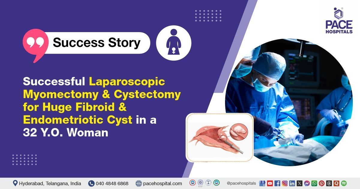
The Gynaecology team at PACE Hospitals successfully performed a laparoscopic myomectomy combined with a left ovarian cystectomy to remove a chocolate cyst and performed morcellation for a 32-year-old female patient who was diagnosed with a large uterine fibroid and a left ovarian endometriotic cyst.
Chief Complaints
A 32-year-old female patient presented to the Gynaecology Department at
PACE Hospitals, Hitech City, Hyderabad, with complaints of menorrhagia, involving abnormally heavy and prolonged menstrual bleeding, along with abdominal pain.
Past History
She had no history of
hypertension,
diabetes mellitus,
hypothyroidism, or
asthma. There was no significant family history, and she reported no known drug or food allergies. Despite the absence of notable medical or familial risk factors, her menstrual and obstetric history revealed regular cycles with consistently heavy bleeding, further supporting the diagnosis of menorrhagia.
General examination
Upon admission to PACE Hospitals, the patient's vital signs were stable. On general examination, the patient was found to be conscious, coherent, and cooperative, with no signs of pallor, icterus, or cyanosis. Cardiovascular examination revealed that S1 and S2 heart sounds were audible, and respiratory system evaluation showed equal air entry bilaterally. On examining the abdomen, the doctor found that the patient’s uterus was enlarged, about the size it would be at 24 weeks of pregnancy, but there was no pain when touched. During the internal examination using a speculum and bimanual examination (per vaginal), the cervix appeared to be pulled upward and had a very small opening.
Diagnosis
After being admitted to PACE Hospitals, the patient underwent a thorough review of her medical history and clinical examination by the gynaecology team, which led to the suspicion of a uterine fibroid and a left ovarian cyst (endometriomas). The diagnosis was further confirmed through imaging studies, including a pelvic ultrasound and an MRI, which provided detailed visualization of the uterine fibroids and the characteristic features of the endometriotic cyst. These diagnostic tools supported the clinical findings and helped guide the treatment plan.
Based on these findings, further diagnostic evaluation was conducted, and she was advised to undergo Endometriotic cyst / endometrioma and Uterine Fibroid Treatment in Hyderabad, India, under the expert care of the Gynaecology Department, ensuring comprehensive evaluation and management tailored to the patient's needs.
Medical Decision-Making
Following consultation with Dr. Mugdha Bandawar, the consultant gynaecologist/gynaecology doctor, and other specialists, including Dr. Mounika Jetti, a comprehensive evaluation was conducted to determine the best diagnostic and therapeutic approach for the patient.
Based on the clinical findings and expert assessment, it was decided that a laparoscopic myomectomy surgery combined with a left ovarian cystectomy for the chocolate cyst and morcellation would be the most effective treatment. This minimally invasive procedure was selected to address the patient’s symptoms while allowing for accurate diagnosis and treatment of the underlying condition.
Surgical Procedure
Following the clinical suspicion, the patient was scheduled to undergo a Laparoscopic Myomectomy in Hyderabad at PACE Hospitals, combined with a left ovarian cystectomy for a chocolate cyst, along with morcellation under the expert supervision of the Gynaecology Department.
Laparoscopic myomectomy combined with left ovarian cystectomy is a minimally invasive surgical procedure designed to treat the simultaneous presence of uterine fibroids and ovarian endometriotic cysts. This technique allows for the removal of non-cancerous fibroids while preserving the uterus, and simultaneously excises the endometriotic (chocolate) cyst from the ovary. Morcellation is used to fragment the fibroids into smaller pieces, enabling their removal through small laparoscopic incisions. This advanced approach effectively relieves symptoms, promotes faster recovery, minimizes scarring, and significantly reduces postoperative pain and discomfort.
Intraoperative Findings
Doctors found an 18 x 15 cm subserosal fibroid arising from the posterior wall and fundus of the uterus, occupying the entire pelvic cavity and causing significant distortion of the pelvic anatomy. Subserosal fibroids of this size can lead to symptoms such as abdominal distension, pelvic pressure, urinary frequency, and discomfort.
Additionally, the uterine wall contained two to three smaller intramural fibroids, the largest measuring approximately 3 x 3 cm. Intramural fibroids, located within the uterine musculature, are known to cause symptoms such as heavy menstrual bleeding, pelvic pain, and potential fertility issues, depending on their size and location.
Both fallopian tubes appeared normal on examination, with no evidence of abnormalities or tubal blockage noted bilaterally.
The right ovary was normal in size, shape, and structure, with no visible cysts, masses, or other abnormalities. However, a 4 x 4 cm endometriotic cyst (chocolate cyst) was identified on the left ovary. A cystectomy was performed to remove the cyst, with careful preservation of the surrounding healthy ovarian tissue.
The uterine fibroids were surgically enucleated, preserving the integrity of the surrounding uterine tissue. The myoma bed was subsequently sutured using the baseball stitch technique to ensure proper closure and promote optimal healing.
Intraoperatively, one unit of packed red blood cells (PRBC) was transfused to manage blood loss and maintain adequate haemoglobin levels.
Morcellation was employed to facilitate the removal of the large fibroid. This technique involves fragmenting the fibroid into smaller pieces to enable extraction, particularly beneficial in minimally invasive surgical approaches.
Postoperative Care
Postoperatively, the patient was monitored in the Surgical Intensive Care Unit (SICU), where she received intravenous (IV) antibiotics, intravenous (IV) pain relief, and intravenous (IV) fluids for supportive care. Due to a postoperative drop in hemoglobin levels, one unit of packed red blood cells (PRBC) was transfused. Once the patient was stabilized, she was transferred to the general ward. Her postoperative course remained uneventful, and a follow-up ultrasound screening was performed, which showed normal findings.
Discharge notes
The patient was discharged in a stable condition.
Medications Advised at the stay
During the hospital stay, the patient was managed with medications including antibiotics, antiemetics, antifibrinolytics, analgesics, proton pump inhibitors (PPIs), and nonsteroidal anti-inflammatory drugs (NSAIDs).
Discharge Medications
Upon discharge, the patient was prescribed a combination of medications, including oral antibiotics, proton pump inhibitors (PPIs), analgesics for pain management, and a topical antibiotic ointment for local application for 15 days. The patient was also advised to maintain a soft diet during the recovery period.
Advice on Discharge
The patient was advised to avoid heavy lifting and strenuous physical activity during the recovery period to promote optimal healing. Additionally, it was recommended to defer any pregnancy planning for at least 9 to 12 months post-surgery, allowing sufficient time for complete physical recovery.
Emergency Care
The patient was instructed to seek immediate medical attention at the Emergency ward of PACE Hospitals in case of any alarming symptoms, including fever, abdominal pain, diarrhoea, constipation, or any other signs of a potential medical emergency.
Review and follow-up notes
The patient was advised to return for a follow-up appointment with the Gynecologist at PACE Hospitals in Hyderabad after 10 days. She was instructed to bring the biopsy report along and to ensure that a prior appointment was scheduled for the visit.
Conclusion
This case highlights the effectiveness of minimally invasive surgical techniques in managing complex gynaecological conditions, such as huge fibroid uterus with a left ovarian endometriotic cyst. A successful laparoscopic myomectomy, combined with a left ovarian cystectomy, was performed to treat large uterine fibroid and endometriotic (chocolate) cyst. The procedure involved the meticulous removal of multiple fibroids from the uterus and an endometriotic cyst from the left ovary. To facilitate extraction, morcellation was employed to break down the larger fibroid into smaller, manageable fragments.
The Role of Pelvic Ultrasound in Diagnosing Gynaecological Conditions
Pelvic ultrasound plays a vital role in the diagnosis and management of gynaecological conditions such as uterine fibroids and ovarian cysts. In this case, the ultrasound findings reveal a large fibroid in the uterus, characterized by abnormal growth of the muscle tissue, and a left ovarian endometriotic cyst (commonly known as a chocolate cyst), which develops when endometrial tissue grows outside the uterus. These conditions are often associated with symptoms such as pelvic pain, heavy menstrual bleeding, and fertility issues. The imaging results provide essential information for accurately identifying the size, location, and characteristics of these lesions, which is crucial for formulating an effective and individualized treatment plan.
Share on
Request an appointment
Fill in the appointment form or call us instantly to book a confirmed appointment with our super specialist at 04048486868
Appointment request - health articles